. 2025 May 1;16(1):4090.
doi: 10.1038/s41467-025-58889-x.

Filamin C dimerisation is regulated by HSPB7

Affiliations

Filamin C dimerisation is regulated by HSPB7

Zihao Wang et al. Nat Commun. .

Abstract

The biomechanical properties and responses of tissues underpin a variety important of physiological functions and pathologies. In striated muscle, the actin-binding protein filamin C (FLNC) is a key protein whose variants causative for a wide range of cardiomyopathies and musculoskeletal pathologies. FLNC is a multi-functional protein that interacts with a variety of partners, however, how it is regulated at the molecular level is not well understood. Here we investigate its interaction with HSPB7, a cardiac-specific molecular chaperone whose absence is embryonically lethal. We find that FLNC and HSPB7 interact in cardiac tissue under biomechanical stress, forming a strong hetero-dimer whose structure we solve by X-ray crystallography. Our quantitative analyses show that the hetero-dimer out-competes the FLNC homo-dimer interface, potentially acting to abrogate the ability of the protein to cross-link the actin cytoskeleton, and to enhance its diffusive mobility. We show that phosphorylation of FLNC at threonine 2677, located at the dimer interface and associated with cardiac stress, acts to favour the homo-dimer. Conversely, phosphorylation at tyrosine 2683, also at the dimer interface, has the opposite effect and shifts the equilibrium towards the hetero-dimer. Evolutionary analysis and ancestral sequence reconstruction reveals this interaction and its mechanisms of regulation to date around the time primitive hearts evolved in chordates. Our work therefore shows, structurally, how HSPB7 acts as a specific molecular chaperone that regulates FLNC dimerisation.


Conflict of interest statement

Competing interests: The authors declare no competing interests

Figures

Fig. 1
Fig. 1. HSPB7 and FLNC are up-regulated and interact in biomechanical stress models in mouse.
A Co-immunoprecipitation of HSPB7 and FLNC from MLP KO mouse ventricular tissue. The co-precipitation of the two proteins when precipitating FLNC suggests they form (at least part of) a complex. An isotype control, where a non-specific antibody (rather than for FLNC) raised in the same species was used as a negative control and showed no bands, whereas a positive control using an aliquot of lysate showed clear bands for HSPB7. Molecular mass bands are marked, in units of kDa. One biological repeat, immunoprecipitation experiment performed in triplicate, one representative shown. B Western blots for FLNC and HSPB7 in MLP KO, TAC and IsoPE mouse ventricular tissue. GAPDH was used as the loading control. Sham and saline are the respective specific negative controls for TAC and IsoPE. In all stress treatments, HSPB7 and FLNC are observed at higher levels. Molecular mass bands are marked, in units of kDa. Two biological replicates (i.e. two mice), blots repeated twice, one representative blot shown. C Immunofluorescence of HSPB7 and FLNC from frozen sections of ventricular tissues from TAC, IsoPE and MLP KO mouse models. Sections were stained for FLNC (Green) and HSPB7 (Red). In all stress treatments, the two proteins are up-regulated and co-localise. For quantification of co-localisation see Supplementary Fig. 1. Scale bars are 25 μm. Two biological replicates (i.e. two mice), two images taken from each sample, one representative image shown.
Fig. 2
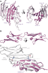
Fig. 2. Crystal structure of the HSPB7 monomeric ACD.
A The asymmetric unit of HSPB7ACDC131S contains three chains (chains A–C), packed through the neighbouring β4 and β6 + 7 strands. B The Ig-fold is very similar to that of other sHSP ACDs: it contains six β strands, with a groove between β4 and β8 and an extended “β6 + 7” strand. C Overlay of a HSPB7ACDC131S (purple) structural view with one of the closely related HSPB1 (white) reveals how the absence in HSPB7 of specific salt bridges (e.g. D129-R140 in HSPB1, highlighted here) that are conserved in the other HSPBs (Supplementary Fig. 10B) have weakened the dimer interface such that monomers are the dominant species in solution.
Fig. 3
Fig. 3. Hetero-dimerisation of HSPB7 and FLNC.
A Native MS experiments show that FLNCd24 (red) forms heterodimers (black) with HSPB7, HSPB7ΔN and HSPB7ACD (purple). No complexes between the FLNCd24 homodimer and HSPB7 are observed, revealing how homo- and hetero-dimerisation are directly competitive. Proteins were mixed at a monomer ratio of 1:1, but the instability of full-length HSPB7 means it was somewhat depleted in solution such that FLNCd24 was effectively in excess (and FLNCd24 is visible in the spectrum). B The FLNCd24:HSPB7ACDC131S heterodimer structure reveals an interface that involves the HSPB7 β4, β8 strands and FLNCd24 strands C and D, centred on the parallel pairing of strands C and β4. C The hydrophobic β4-8 groove on HSPB7 accepts M2667 and M2669 from FLNC (top), while the groove between strands C and D on FLNCd24 accepts I102 from HSPB7 (middle). This leads to a network of hydrophobic interactions that stabilise the heterodimer (bottom). D HDX-MS analysis of HSPB7ACD and FLNCd24 in the heterodimer compared to in isolation. Woods plots showing the difference in deuterium uptake of HSPB7ACD (top) and FLNCd24 (bottom) in the two states, at a labelling time of 500 s. The y-axis is calculated as the uptake for the isolated proteins minus that in the heterodimer; negative values denote protection from exchange in the heterodimer. Three technical repeats were carried out, and peptides were considered significantly different if p < 0.01 (coloured: orange or purple). E Plot of deuterium uptake versus exposure time of the most protected peptide in HSPB7ACD (upper) and FLNCd24 (lower) in the heterodimer compared to this same peptide in the isolated protein. Because HSPB7 is monomeric, in the absence of FLNC, this peptide is solvent-exposed and therefore has consistently high uptake values. Error bars refer to the standard deviation of three repeats at each time-point, and the shaded bands 99% confidence. F Mapping the uptake difference at 500 s (as a fraction of the theoretical maximum) onto the heterodimer protein structure shows that protection is centred on the dimer interface that we found in our crystal structure.
Fig. 4
Fig. 4. Mimicking phosphorylation at T2677 and Y2683 has opposing effects in modulating FLNCd24 homo- and hetero-dimerisation.
A Native mass spectra at 1 μM of FLNCd24 WT (middle) and the phosphomimics T2677D (left) and Y2683E (right) show differences in the relative abundances of the monomer (yellow) and dimer (red) (upper row). T2677D forms a stabilised homodimer, relative to WT; Y2683 a destabilised dimer. Native MS spectra at 5 μM of each of HSPB7ACD and either FLNCd24 WT or one of the phosphomimics (lower row). Heterodimers are the dominant species in each of the spectra, but a notable abundance of FLNC (red, and yellow) and HSPB7ACD (purple) can be observed in the case of the T2677D mutant, and to a lesser extent for WT and Y2683E. B Titration curves for homo- (upper) and hetero-dimerisation (lower). WT: red and medium grey; T2677D: salmon and light grey; Y2683D: burgundy and dark grey. Error bars represent ±1 standard deviation of the mean (n = 3). C Free energy diagram showing the differences in stability for homo- and heterodimers in each of the three forms of FLNC. The vertical thickness of the bars corresponds to the error in our estimates. Lower free energies correspond to higher stability. See also Supplementary Methods.
Fig. 5
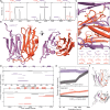
Fig. 5. MD simulations reveal how phosphorylation modulates the strength of the FLNCd24 homo- and heterodimer interfaces.
A The location of the T2677 and Y2683 phosphorylation sites on the FLNCd24 homodimer (PDB 1V05) and FLNCd24:HSPB7ACDC131S heterodimer structures is near the interface. FLNC: orange; HSPB7: purple. B Difference map of hydrogen bond occupancy between WT and either pT2677 (lower right triangle) or pY2683 FLNCd24 (upper left) homodimers. Each pixel corresponds to a particular inter-monomer contact between residue pairs. Blue: hydrogen bond formed more in the phosphorylated form; red: hydrogen bond formed more in the WT. A highly occupied hydrogen bond formed across the WT homodimer interface (between E2681 and Y2683) is lost upon phosphorylation of Y2683 (WT 71%; pY2683 0%). And a new interaction with R2690 is formed upon phosphorylation of T2677 (WT 0%; pT2677 26%) (insets). C Difference maps of hydrogen bond occupancy between WT and pT2677 (upper) or pY2683 FLNCd24:HSPB7ACDC131S heterodimers (lower). Colouring as in (B). The most substantial differences are highlighted with representative frames from the trajectories showing the contacts made (insets). These are: a highly occupied hydrogen bond formed across the WT heterodimer interface (between G2674 and N107) lost upon phosphorylation of T2677 (WT 63%; pT2677 1%); and a new interaction is formed between pY2683 with R113 that is absent in the WT (WT 0%; pY2683 32%). Altered interactions are also observed between residues that do not directly involve the phosphorylated site, e.g. a contact between D2712 and T143 (WT 10%; pT2677 38%) and ones between the cluster H2686/N2689/R2690 and E99 (WT 0/0/22%; pY2683 28/41/45%). For a full inventory of hydrogen bond occupancies, see the Supplementary Data 1.
Fig. 6
Fig. 6. Overlapping interfaces for homo- and hetero-dimerisation lead to an equilibrium that can be regulated by phosphorylation to adjust FLNC mobility.
A Plot of the distances between subunits in the homo- (orange) and heterodimer (black), as a function of sequence position in our MD simulations. The bands are defined by the closest (lower value) and average (upper value) distance over the length of the simulation (upper). The two bands overlap well, demonstrating the overall similarity of the interfaces in the two dimers. The core dimer interface is marked (dashed box), and expanded in the lower panel, which shows the average distance only, but for each of the WT, pT2677 and pY2683 simulations. These graphs show that the dimer interface distances are highly similar across the three proteins, with minor differences found only near pT2677 (for that protein). This is consistent with the overall structure of the dimer interface being largely similar, with the major differences between WT and phosphorylated forms lying in the hydrogen-bonds across the interface (Fig. 5). B Colouring residues on the surface in proportion to the closest distances of a WT FLNC monomer to its counterpart reinforces the similarity in binding site between homo- and heterodimer (insets). This allows us a simple view of the equilibria involved, where FLNCd24 homo- and hetero-dimerisation are competitive. The equilibrium favours the heterodimer for WT FLNC, and can be shifted in either direction by phosphorylation. C The shifting of the equilibrium effectively adjusts the state of FLNC, either stabilising it in the actin-crosslinking-competent homodimer form, or effectively releasing it from this role by generating the much smaller heterodimer, which is likely much more mobile in the cell.

References

    1. Tarone, G. & Brancaccio, M. Keep your heart in shape: molecular chaperone networks for treating heart disease. Cardiovasc. Res.102, 346–361 (2014). - PubMed
    1. Zhou, A. X., Hartwig, J. H. & Akyürek, L. M. Filamins in cell signaling, transcription and organ development. Trends Cell Biol.20, 113–123 (2010). - PubMed
    1. Razinia, Z., Mäkelä, T., Ylänne, J. & Calderwood, D. A. Filamins in mechanosensing and signaling. Annu. Rev. Biophys.41, 227–246 (2012). - PMC - PubMed
    1. Van Der Ven, P. F. M. et al. Characterization of muscle filamin isoforms suggests a possible role of γ-Filamin/ABP-L in sarcomeric Z-disc formation. Cell Motil. Cytoskeleton45, 149–162 (2000). - PubMed
    1. Leber, Y. et al. Filamin C is a highly dynamic protein associated with fast repair of myofibrillar microdamage. Hum. Mol. Genet.25, 2776–2788 (2016). - PubMed