. 2025 May 1;8(1):689.
doi: 10.1038/s42003-025-08130-8.

Mannose inhibits PKM2 lactylation to induce pyroptosis in bladder cancer and activate antitumor immune responses

Affiliations

Mannose inhibits PKM2 lactylation to induce pyroptosis in bladder cancer and activate antitumor immune responses

Haoyi Jin et al. Commun Biol. .

Abstract

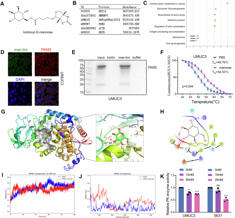
Bladder cancer therapy remains challenging due to poor efficacy and frequent recurrence. Mannose, a naturally occurring monosaccharide, has demonstrated antitumor effects in various cancers, yet its mechanism of action in bladder cancer is unclear. This study explored the inhibitory effects of mannose on bladder cancer. We found mannose significantly inhibited the growth of bladder cancer cells, xenografts, and organoids. Mannose directly binds to PKM2, inhibiting its enzymatic activity and reducing lactate production. This reduction in lactate led to decreased PKM2 lactylation and increased acetylation, causing PKM2 to translocate to the nucleus. Nuclear PKM2 activated the NF-κB pathway, inducing NLRP1/Caspase-1/GSDMD/IL-1β-dependent pyroptosis. Additionally, mannose promoted antitumor immune responses by inducing pyroptosis and enhancing the efficacy of immune checkpoint inhibitors. These findings highlight the use of mannose as a potent antitumor agent and a promising therapeutic strategy for bladder cancer.


Conflict of interest statement

Competing interests: The authors declare no competing interests. Ethics approval and consent to participate: The animal studies were approved by the Animal Care and Use Committee of China Medical University (IACUC Issue No. KT2023184, date: 04/03/2024), and all the animal handling procedures were conducted in accordance with the Basel Declaration. Organoid culture was approved by the local ethics committee of Liaoning Cancer Hospital& Institute (GZR20240373, date: 07/03/2024) based on the Helsinki Declaration, and written informed consent was obtained from all subjects. Consent for publication: All authors consent for publication.

Figures

Fig. 1
Fig. 1. Mannose inhibits tumor growth in bladder cancer.
A Western blot analysis of PMI expression in urinary epithelial cells and bladder cancer cells (SVHUC1, 5637, T24, UMUC3). B Colony formation assay assessing the effects of mannose on the proliferation of urinary epithelial cells (SVHUC1) and cancer cells. C A CCK-8 assay was used to determine the duration of mannose treatment and the IC50 at 48 h, with values of ~45 mM for UMUC3 cells and 20 mM for 5637 cells. D Cell cycle analysis following mannose treatment in UMUC3 and 5637 cells. E PCR analysis of cyclin expression in UMUC3 and 5637 cells treated with mannose. F Flow cytometry analysis of UMUC3 and 5637 cell death induced by mannose treatment. G Assessment of apoptosis pathway activation in mannose-treated UMUC3 and 5637 cells. H Evaluation of the ability of mannose to inhibit the growth of bladder tumor organoids. I Assessment of the growth inhibition effect of mannose in nude mice bearing transplanted bladder tumors. J Immunohistochemistry (Ki-67 staining) confirming the inhibitory effect of mannose on bladder cancer tissue. K Detection of PARP cleavage in cells treated with mannose combined with cisplatin (CDDP) or pirarubicin (THP). L Analysis of the combined effects of mannose and cisplatin or pirarubicin on UMUC3 cells, with drug combination indices calculated via ZIP scores.
Fig. 2
Fig. 2. Mannose binds to PKM2 and inhibits its enzymatic activity.
A Schematic of mannose with biotin modification. B List of proteins bound to biotinylated mannose identified via a pull-down assay and mass spectrometry. C KEGG analysis showing the enrichment of mannose-bound proteins in glycolytic and other metabolic pathways. D Immunofluorescence staining indicating the colocalization of biotinylated mannose with PKM2 in cells. E Confirmation of the binding of mannose to PKM2 via a biotinylated mannose pull-down assay. F CETSA confirming the increased thermal stability of PKM2 in the presence of mannose. G, H Molecular docking results showing that mannose binds to the active pocket of PKM2 and forms hydrogen bonds with the catalytic site K296. I, J Molecular dynamics simulation of PKM2 in complex with mannose, analyzing changes in RMSD and RMSF. K Analysis of the effects of mannose on pyruvate kinase activity in bladder cancer cells.
Fig. 3
Fig. 3. Mannose inhibits lactate production and lactylation.
A Measurement of the pH of the culture medium of bladder cancer cells following mannose treatment. B Seahorse analysis of the impact of mannose on the glycolytic capacity of UMUC3 and 5637 cells. C PLS-DA validation of the effects of mannose on the metabolic profile of UMUC3 bladder cancer cells. D KEGG pathway enrichment analysis of significantly altered metabolic pathways in UMUC3 cells treated with mannose. E Changes in glycolytic products in mannose-treated UMUC3 cells analyzed via metabolomics. F Analysis of LDH enzyme activity in bladder cancer cells treated with mannose. G Measurement of lactate levels in bladder cancer cells following mannose treatment. H Analysis of pyruvate levels in bladder cancer cells treated with mannose. I Assessment of ATP production in mannose-treated bladder cancer cells. J Restoration of ATP production by lactate supplementation in mannose-treated bladder cancer cells. K Western blot analysis of lactylation levels in mannose-treated bladder cancer cells. L Immunohistochemistry was used to assess the effects of mannose on protein lactylation levels in xenograft tumor tissues from nude mice.
Fig. 4
Fig. 4. Mannose induces PKM2 nuclear translocation by regulating K433 lactylation.
A MS identification of K433 on PKM2 as a potential lactylation site in bladder cancer cells. B Immunoprecipitation validation of lactylation of PKM2 identified by mass spectrometry. C Validation of the regulatory effects of mannose on the identified lactylation of PKM2 in bladder cancer cells. D Introduction of exogenous wild-type PKM2 and K433R mutant PKM2 in bladder cancer cells, comparing lactylation levels with those of lactate supplementation. E Comparison of pyruvate production by wild-type PKM2 and K433R mutant PKM2 in bladder cancer cells. F Immunoprecipitation validation of the interaction between PKM2 and p300 in bladder cancer cells. G Changes in PKM2 lactylation levels in bladder cancer cells after p300 was knocked down with two different siRNAs. H Effects of p300 knockdown on pyruvate kinase activity in bladder cancer cells. I Effects of mannose and lactate supplementation on the lactylation and acetylation levels of PKM2 in bladder cancer cells. J Validation of PKM2 phosphorylation in mannose-treated bladder cancer cells. K Fluorescence detection of PKM2 localization following mannose treatment. L Analysis of nuclear and cytoplasmic PKM2 expression in bladder cancer cells. M Examination of the effects of mannose, glucose, and lactate supplementation on nuclear PKM2 expression in bladder cancer cells.
Fig. 5
Fig. 5. Mannose regulates PKM2 nuclear translocation to activate the NF-κB pathway and promote pyroptosis.
A RNA sequencing of mannose-treated UMUC3 cells with transcription factor analysis exploring key transcription factors associated with differentially expressed genes, highlighting the significant associations with NFKB1 and RELA. B Construction of bladder cancer cell lines with nuclear-specific expression of PKM2 to assess the effects of mannose treatment and nuclear PKM2 on the expression levels of p50 and p65. C Immunohistochemistry was used to assess the impact of mannose treatment on NF-κB transcription factor expression in xenograft tumor tissues from nude mice. D Fluorescence colocalization evaluation of p50 or p65 with PKM2 in UMUC3 cells. E Immunoprecipitation of p50 or p65 combined with PKM2 in UMUC3 and 5637 cells. F Analysis of the effects of mannose and lactate on p50 and p65 expression levels in different bladder cancer cell lines. G KEGG pathway enrichment analysis of significantly altered pathways in UMUC3 cells treated with mannose identified through RNA sequencing, highlighting pathways related to pyroptosis. H Measurement of LDH release induced by mannose-induced pyroptosis in bladder cancer cells. I Analysis of lipid peroxidation levels in bladder cancer cells treated with mannose by measuring the MDA content. J Examination of morphological changes in UMUC3 and 5637 cells via scanning electron microscopy (SEM) following mannose treatment.
Fig. 6
Fig. 6. Mannose activates NLRP1/Caspase-1/GSDMD/IL-1β-dependent pyroptosis.
A PCR analysis of NF-κB-induced cytokine expression in mannose-treated bladder cancer cells. B PCR analysis of pyroptosis-induced gasdermin gene expression in mannose-treated bladder cancer cells. C PCR analysis of genes upstream of GSDMD in mannose-treated bladder cancer cells. D Examination of the activation of pyroptosis pathways in response to mannose in bladder cancer cells. E Examination of the activation of NOD-like receptor pathways in response to mannose in bladder cancer cells. F Examination of the activation of NLRP1 and Caspase-1 by supplementation with lactate in mannose-treated bladder cancer cells. G Examination of LDH release by the addition of lactate to mannose-treated bladder cancer cells. Analysis of the transcriptional regulation of pyroptosis genes by the NF-κB transcription factor p65: H IL-1β, I GSDMD, J Caspase-1, K NLRP1. L. Immunohistochemistry was used to assess the impact of mannose treatment on pyroptosis-related protein expression levels in xenograft tumor tissues from nude mice.
Fig. 7
Fig. 7. Mannose activates antitumor immune responses through pyroptosis.
A Impact of mannose feeding on the growth of MB49 subcutaneous transplant tumors in C57BL/6 mice on day 23. B Tumor growth curves of the mannose-supplemented subcutaneous transplant tumors. C Effects of mannose supplementation on the body weights of C57BL/6 mice on day 23. D Effects of mannose feeding on the blood glucose levels of C57BL/6 mice on day 23. E Immune infiltration analysis in mannose-supplemented transplant tumors harvested on day 23 via RNA sequencing, with a heatmap showing the fold changes in immune cell infiltration scores between the two groups. F IHC was used to assess the impact of mannose exposure on the infiltration levels of CD8+ T cells, cytotoxic CD8+ T cells (Gzmb), and CD11b+ innate immune cells, and Pd-l1 expression in mouse tumors harvested on day 23. G PCR analysis of immune-related interleukins in mouse tumors harvested on day 23. H Fluorescence evaluation of GSDMD expression in mouse tumors harvested on day 23. I Effects of the combination of mannose and an anti-Pd-1 antibody on the growth of MB49 subcutaneous transplant tumors in C57BL/6 mice. J Tumor growth curves of subcutaneously transplanted tumors treated with mannose combined with an anti-Pd-1 antibody.

References

    1. Siegel, R. L., Miller, K. D., Wagle, N. S. & Jemal, A. Cancer statistics, 2023. CA Cancer J. Clin.73, 17–48 (2023). - PubMed
    1. Seiler, R. et al. Impact of molecular subtypes in muscle-invasive bladder cancer on predicting response and survival after neoadjuvant chemotherapy. Eur. Urol.72, 544–554 (2017). - PubMed
    1. Liu, S., Chen, X. & Lin, T. Emerging strategies for the improvement of chemotherapy in bladder cancer: Current knowledge and future perspectives. J. Adv. Res.39, 187–202 (2022). - PMC - PubMed
    1. Facchini, G. et al. Advanced/metastatic bladder cancer: current status and future directions. Eur. Rev. Med. Pharmacol. Sci.24, 11536–11552 (2020). - PubMed
    1. Watkins, C. B. & Frenkel, C. Inhibition of pear fruit ripening by mannose. Plant Physiol.85, 56–61 (1987). - PMC - PubMed

MeSH terms