

A deep learning algorithm for automated adrenal gland segmentation on non-contrast CT images
- PMID: 40312690
- PMCID: PMC12046700
- DOI: 10.1186/s12880-025-01682-5
A deep learning algorithm for automated adrenal gland segmentation on non-contrast CT images
Abstract
Background: The adrenal glands are small retroperitoneal organs, few reference standards exist for adrenal CT measurements in clinical practice. This study aims to develop a deep learning (DL) model for automated adrenal gland segmentation on non-contrast CT images, and to conduct a preliminary large-scale study on age-related volume changes in normal adrenal glands using the model output values.
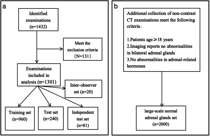
Methods: The model was trained and evaluated on a development dataset of annotated non-contrast CT scans of bilateral adrenal glands, utilizing nnU-Net for segmentation task. The ground truth was manually established by two experienced radiologists, and the model performance was assessed using the Dice similarity coefficient (DSC). Additionally, five radiologists provided annotations on a subset of 20 randomly selected cases to measure inter-observer variability. Following validation, the model was applied to a large-scale normal adrenal glands dataset to segment adrenal glands.
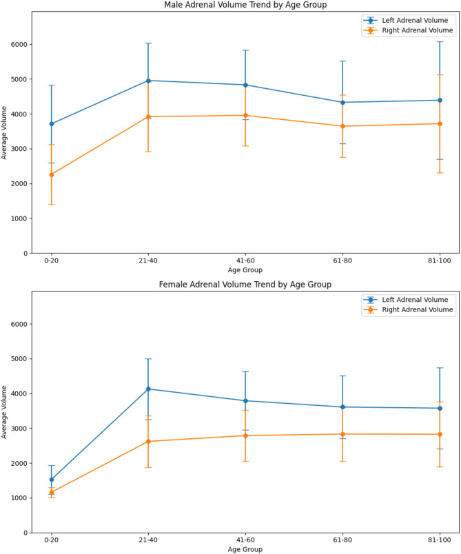
Results: The DL model development dataset contained 1301 CT examinations. In the test set, the median DSC scores for the segmentation model of left and right adrenal glands were 0.899 and 0.904 respectively, and in the independent test set were 0.900 and 0.896. Inter-observer DSC for radiologist manual segmentation did not differ from automated machine segmentation (P = 0.541). The large-scale normal adrenal glands dataset contained 2000 CT examinations, the graph shows that adrenal gland volume increases first and then decreases with age.
Conclusion: The developed DL model demonstrates accurate adrenal gland segmentation, and enables a comprehensive study of age-related adrenal gland volume variations.
Keywords: Adrenal gland; Adrenal gland segmentation; Dice similarity coefficient; Non-contrast CT; nnU-Net.
© 2025. The Author(s).
Conflict of interest statement
Declarations. Ethics approval and consent to participate: This study was approved by the Ethics Committee of Fuwai Central-China Cardiovascular Hospital (Approval No. 2025.21). Given the retrospective nature of the study and the use of fully anonymized patient data, the requirement for informed consent was waived by the ethics committee. All procedures were conducted in accordance with the ethical standards outlined in the Declaration of Helsinki and its later amendments. Consent for publication: Not applicable. Competing interests: The authors declare no competing interests.
Figures


Similar articles
-
Machine Learning for Adrenal Gland Segmentation and Classification of Normal and Adrenal Masses at CT.Radiology. 2023 Feb;306(2):e220101. doi: 10.1148/radiol.220101. Epub 2022 Sep 20. Radiology. 2023. PMID: 36125375
-
Fully automatic volume measurement of the adrenal gland on CT using deep learning to classify adrenal hyperplasia.Eur Radiol. 2023 Jun;33(6):4292-4302. doi: 10.1007/s00330-022-09347-5. Epub 2022 Dec 26. Eur Radiol. 2023. PMID: 36571602
-
nnU-Net-Based Pancreas Segmentation and Volume Measurement on CT Imaging in Patients with Pancreatic Cancer.Acad Radiol. 2024 Jul;31(7):2784-2794. doi: 10.1016/j.acra.2024.01.004. Epub 2024 Feb 12. Acad Radiol. 2024. PMID: 38350812
-
Adrenal Volume Quantitative Visualization Tool by Multiple Parameters and an nnU-Net Deep Learning Automatic Segmentation Model.J Imaging Inform Med. 2025 Feb;38(1):47-59. doi: 10.1007/s10278-024-01158-y. Epub 2024 Jul 2. J Imaging Inform Med. 2025. PMID: 38955963 Free PMC article.
-
Unleashing the strengths of unlabelled data in deep learning-assisted pan-cancer abdominal organ quantification: the FLARE22 challenge.Lancet Digit Health. 2024 Nov;6(11):e815-e826. doi: 10.1016/S2589-7500(24)00154-7. Lancet Digit Health. 2024. PMID: 39455194 Review.
References
-
- Navin PJ, Moynagh MR. Optimal and novel imaging of the adrenal glands. Curr Opin Endocrinol Diabetes Obes. 2022;29(3):253–62. 10.1097/MED.0000000000000730. - PubMed
-
- Ctvrtlik F, Koranda P, Tichy T. Adrenal disease: a clinical update and overview of imaging. A review. Biomed Pap Med Fac Univ Palacky Olomouc Czech Repub. 2014;158(1):23–34. 10.5507/bp.2014.010. - PubMed
-
- Mohan DR, Lerario AM. Closing the loop on adrenal health, dysfunction, and disease. Sci Transl Med. 2023;15(701):eadh4450. 10.1126/scitranslmed.adh4450. - PubMed
MeSH terms
Grants and funding
LinkOut - more resources
Full Text Sources
Medical
