. 2025 May 1;23(1):500.
doi: 10.1186/s12967-025-06229-4.

LncRNA PVT1 activated by TGF-β1/Smad3 facilitates proliferation and metastasis of hepatocellular carcinoma via upregulating Smad6 and NRG1

Affiliations

LncRNA PVT1 activated by TGF-β1/Smad3 facilitates proliferation and metastasis of hepatocellular carcinoma via upregulating Smad6 and NRG1

Shuaihui Wu et al. J Transl Med. .

Abstract

Background: Hepatocellular carcinoma (HCC) significantly affects the patient's physical and mental health. Long non-coding RNA plasmacytoma variant translocation 1 (lncRNA PVT1) has been associated with the progression of HCC. However, the current effectiveness of HCC treatment is considered insufficient, and the scope of its therapeutic targets is highly limited. The purpose of this investigation is to investigate the pathogenic mechanism of PVT1 in HCC and assess its potential for gene therapy in HCC.

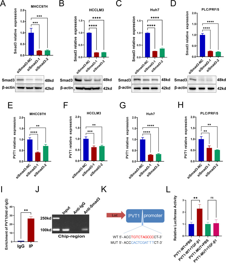
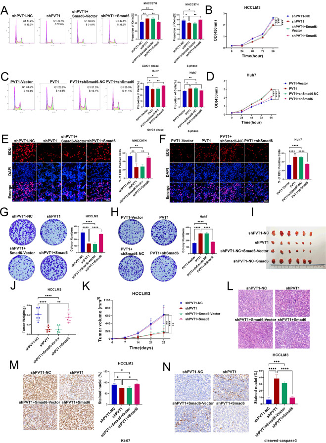
Methods: This study assessed cycle phases and proliferative capacity of HCC cells through flow cytometry, CCK-8 assay, EdU, and colony formation assays. Chromatin Immunoprecipitation (ChIP) and Dual-Luciferase Reporter Assays were conducted to investigate the interactions among the promoter and PVT1, PVT1 and its target miRNAs, as well as miRNAs and their target genes. BALB/c nude mice were employed to establish models for studying the proliferation and metastasis of HCC in vivo.

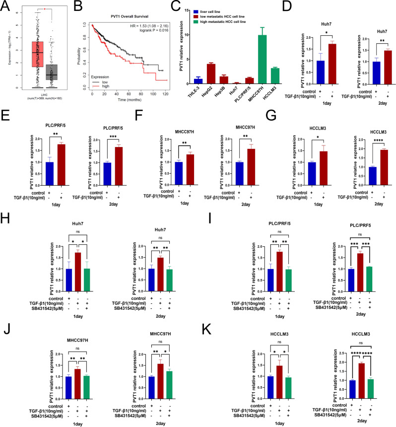
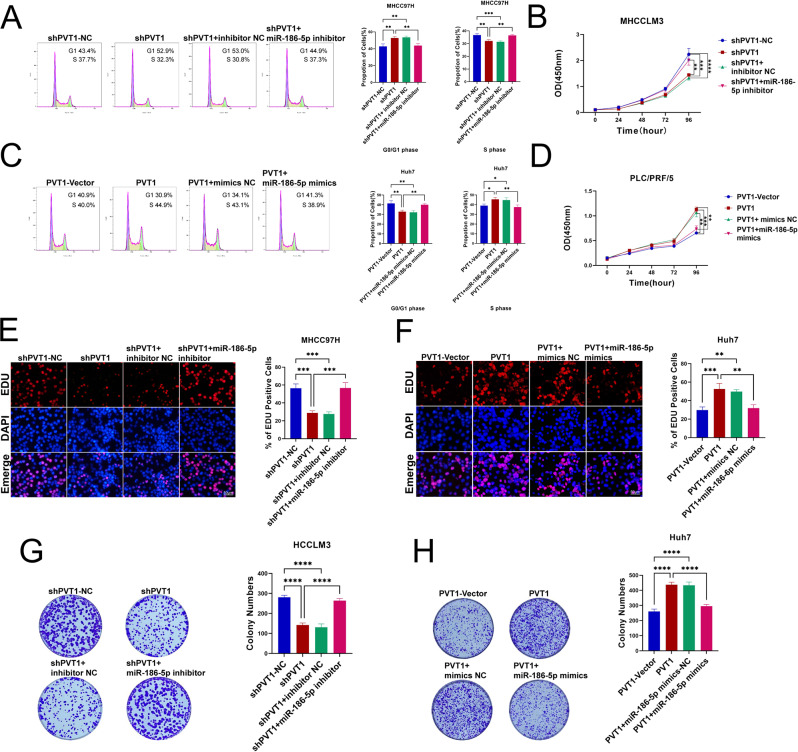
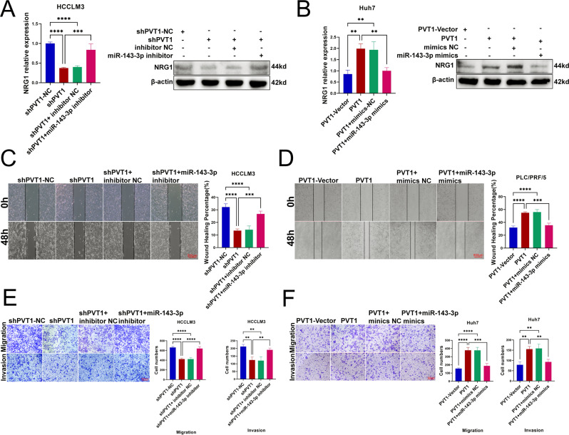
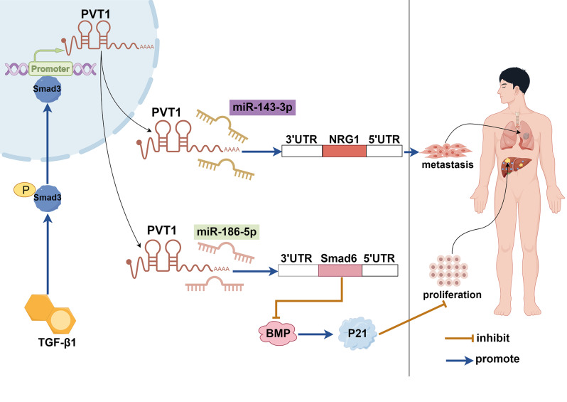
Results: The data revealed that TGF-β1 upregulates PVT1, while Smad3 functions as a transcription factor to modulate PVT1. PVT1, in turn, upregulates Smad6 and NRG1 (Neuregulin 1). Moreover, PVT1 combines with miR-186-5p and miR-143-3p, while miR-186-5p inhibits Smad6 and miR-143-3p inhibits NRG1. Further, in vivo and in vitro analyses revealed that PVT1 stimulates the expression of Smad6, thereby promoting the proliferation of HCC. In addition, PVT1 also promotes the spread of HCC by upregulating NRG1.

Conclusion: This study validated that PVT1 activated by TGF-β1/Smad3 facilitates HCC progression and metastasis by upregulating the miR-186-5p/Smad6 and miR-143-3p/NRG1 axes, indicating its potential as a biological target for treating HCC.

Keywords: Hepatocellular carcinoma; NRG1; P21; Smad3; Smad6; TGF-β1; lncRNA PVT1; miR-143-3p; miR-186-5p.


Conflict of interest statement

Declarations. Ethics approval and consent to participate: All animal experiments in this study were approved by the Ethical Committee of the Zhongnan Hospital of Wuhan University. The in vivo experiments were performed by investigators who had acquired Hubei Province’s Experimental Animal Professional and Technical Certificate. Consent for publication: Not applicable. Competing interests: The authors declare that they have no competing interests.

Figures

Fig. 1
Fig. 1
PVT1 was highly expressed in HCC and up-regulated by TGF-β1. (A) The GEPIA database indicated the PVT1 expression in liver cancer and normal tissue. (B) The Kaplan-Meier Plotter platform assessed the overall survival (OS) in HCC patients. (C) qRT-PCR was carried out to assess the expression of PVT1 in hepatocytes and hepatoma cells. (D-G) qRT-PCR was carried out to assess the expression of PVT1 in Huh7, PLC/PRF/5, MHCC97H, and HCCLM3 cells after treatment with TGF-β1 (10 ng/mL) for 1 and 2 days. (H-K) qRT-PCR was carried out to assess the PVT1 expression after the treatment of SB431542 (5 µM). The data are expressed as the mean ± SD (n = 3). *p < 0.05, **p < 0.01, ***p < 0.001, ****p < 0.0001
Fig. 2
Fig. 2
Smad3 binds to PVT1 and positively regulates its expression as a transcription factor. (A-D) qRT-PCR and WB analyses were performed to assess the knockdown efficiency of Smad3 in MHCC97H, HCCLM3, Huh7, and PLC/PRF/5 cells. (E-H) qRT-PCR data confirmed that PVT1 expression was decreased in four HCC cell types after Smad3 knockdown. (I, J) The ChiP experiment results confirmed the combination of Smad3 and PVT1. (K) The binding and mutation sites of Smad3 and PVT1. (L) The result of dual-luciferase reporter assay in HCCLM3 cells. *p < 0.05, **p < 0.01, ***p < 0.001, ****p < 0.0001
Fig. 3
Fig. 3
PVT1 promotes the proliferation, migration, and invasion of HCC cells. (A, H) qRT-PCR was performed to assess the knockdown efficiency of PVT1 in HCCLM3 cells and the overexpression efficiency in huh7 cells. (B, I) Flow cytometry was conducted to assess the cell cycles of HCCLM3 cells (PVT1 knockdown) and huh7 cells (PVT1 overexpression). (C-E, J-L) The CCK-8, EdU, and colony formation analyses were performed to measure the proliferation of HCCLM3 and huh7 cells after PVT1 knockdown and overexpression. (F, G, M, N) Wound healing and transwell tests were conducted to evaluate the migration and invasion ability of HCCLM3 and huh7 cells. *p < 0.05, **p < 0.01, ***p < 0.001, ****p < 0.0001
Fig. 4
Fig. 4
The downstream target of PVT1. (A, B) qRT-PCR and WB analyses were performed to assess the Smad6 and P21 expression in HCCLM3 (PVT1 knockdown) and huh7 (PVT1 overexpression) cells. (C, D) qRT-PCR and WB analyses were performed to assess NRG1 expression in HCCLM3 (PVT1 knockdown) and huh7 (PVT1 overexpression) cells. (E) Bioinformatics predicted miRNAs binding with PVT1 and Smad6. (F) The miRNAs binding to PVT1 and NRG1 were identified via a biometrics database. (G) miR-186-5p mimics transfection efficiency was assessed using qRT-PCR. (H) qRT-PCR and WB analyses were performed to assess Smad6 expression after miR-186-5p mimics transfection. (I) miR-186-5p inhibitor transfection efficiency was detected by qRT-PCR. (J) qRT-PCR and WB analyzed analyses were performed to assess Smad6 expression after miR-186-5p inhibitor transfection. (K) The miR-143-3p mimics’ transfection was verified by qRT-PCR. (L) NRG1 expression was examined by WB and qRT-PCR after miR-143-3p mimics transfection (M, N) The transfection efficiency of the miR-143-3p inhibitor was detected by qRT-PCR, and the expression of NRG1 was analyzed via WB and qRT-PCR. (O-P) The results of luciferase reporter assay of PVT1/miR-186-5p/Smad6 interaction. (Q-R) The results of reporter gene assays of PVT1/miR-143-3p/NRG1. *p < 0.05, **p < 0.01, ***p < 0.001, ****p < 0.0001
Fig. 5
Fig. 5
Smad6 deletion reversed the proliferative effect of PVT1 on HCC. (A, B, E, G) Flow cytometry, CCK-8, EdU, and colony formation analyses were carried out to assess the proliferation potential of HCC cells when co-transfected with shPVT1 and Smad6. (C, D, F, H) The growth ability of PVT1 overexpression and shSmad6 in HCC cells was evaluated by Flow cytometry, CCK-8, EdU, and colony formation assays. (I) Representative images of mice’s axillary subcutaneous tumors after four weeks of HCC cell injection. (J, K) The tumor’s weight and volume were recorded and analyzed. (L) H&E staining of the tumor. (M, N) Immunohistochemical staining with Ki67 and cleaved-caspase3 markers was employed to assess the tumor’s proliferative potential. *p < 0.05, **p < 0.01, ***p < 0.001, ****p < 0.0001
Fig. 6
Fig. 6
Silencing of NRG1 rescued PVT1-promoted migration and invasion activities of HCC. (A) qRT-PCR test and WB analyses showed the level of NRG1 overexpression. (B) The qRT-PCR and WB techniques were employed to measure NRG1’s expression in shPVT1 and the overexpression of NRG1 in HCC cells. (C) The silencing efficiency of NRG1 was measured by qRT-PCR and WB assays. (D) qRT-PCR and WB analyses were performed to assess the expression of NRG1 in PVT1 overexpression and shNRG1 conditions. (E, G) Wound healing and transwell tests analyzed the metastatic potential of HCC cells with shPVT1 and NRG1 overexpression. (F, H) The ability of HCC cells to migrate and invade after PVT1 overexpression and NRG1 knockdown was estimated by transwell and wound healing assays. (I) Mouse lungs were subjected to H&E staining, and the lung nodules were enumerated. *p < 0.05, **p < 0.01, ***p < 0.001, ****p < 0.0001
Fig. 7
Fig. 7
PVT1 up-regulates Smad6 by competitive binding miR-186-5p to facilitate HCC proliferation. (A, B, E, G) The proliferation of HCC cells after PVT1 knockdown and transfection with miR-186-5p inhibitor was investigated using flow cytometry, CCK-8, EdU, and colony formation assays. (C, D, F, H) HCC cells overexpressing PVT1 and harboring transfected miR-186-5p mimics were evaluated for their growth potential via flow cytometry, CCK-8, EdU, and colony formation assays. *p < 0.05, **p < 0.01, ***p < 0.001, ****p < 0.0001
Fig. 8
Fig. 8
PVT1 up-regulates NRG1 by competitively binding with miR-143-3p to promote the migration and invasion of HCC. (A) qRT-PCR and WB assays were performed to analyze NRG1 expression in HCC cells after co-transfection with shPVT1 and a miR-143-3p inhibitor. (B) qRT-PCR and WB analyses were performed to assess NRG1 expression levels in HCC cells that overexpressed PVT1 and had been transfected with miR-143-3p mimics. (C, E) Wound healing and transwell experiments were conducted to evaluate the migratory and invasive potentials of HCC cells transfected with shPVT1 and miR-143-3p inhibitor (D, F) After the miR-143-3p mimics and PVT1 were transfected into HCC cells, wound healing and transwell assays performed. *p < 0.05, **p < 0.01, ***p < 0.001, ****p < 0.0001
Fig. 9
Fig. 9
The mechanism of PVT1 regulating proliferation and metastasis in HCC

Similar articles

References

    1. Torre LA, Bray F, Siegel RL, Ferlay J, Lortet-Tieulent J, Jemal A. Global cancer statistics, 2012. CA Cancer J Clin. 2015;65:87–108. - PubMed
    1. Shah M, Sarkar D. HCC-Related lncRNAs: roles and mechanisms. Int J Mol Sci. 2024;25. - PMC - PubMed
    1. Kulik L, El-Serag HB. Epidemiology and management of Hepatocellular Carcinoma. Gastroenterology. 2019;156:477– 91.e1. - PMC - PubMed
    1. Martínez-Barriocanal Á, Arango D, Dopeso H. PVT1 long non-coding RNA in gastrointestinal Cancer. Front Oncol. 2020;10:38. - PMC - PubMed
    1. Zhou C, Yi C, Yi Y, Qin W, Yan Y, Dong X, et al. LncRNA PVT1 promotes gemcitabine resistance of pancreatic cancer via activating Wnt/β-catenin and autophagy pathway through modulating the miR-619-5p/Pygo2 and miR-619-5p/ATG14 axes. Mol Cancer. 2020;19:118. - PMC - PubMed

MeSH terms

LinkOut - more resources