. 2026 Apr 1;21(4):1409-1427.
doi: 10.4103/NRR.NRR-D-24-01507. Epub 2025 Apr 29.

Inherent potential of mitochondria-targeted interventions for chronic neurodegenerative diseases

Affiliations

Inherent potential of mitochondria-targeted interventions for chronic neurodegenerative diseases

Min Zhou et al. Neural Regen Res. .

Abstract

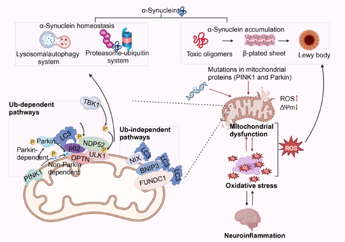
The cure rate for chronic neurodegenerative diseases remains low, creating an urgent need for improved intervention methods. Recent studies have shown that enhancing mitochondrial function can mitigate the effects of these diseases. This paper comprehensively reviews the relationship between mitochondrial dysfunction and chronic neurodegenerative diseases, aiming to uncover the potential use of targeted mitochondrial interventions as viable therapeutic options. We detail five targeted mitochondrial intervention strategies for chronic neurodegenerative diseases that act by promoting mitophagy, inhibiting mitochondrial fission, enhancing mitochondrial biogenesis, applying mitochondria-targeting antioxidants, and transplanting mitochondria. Each method has unique advantages and potential limitations, making them suitable for various therapeutic situations. Therapies that promote mitophagy or inhibit mitochondrial fission could be particularly effective in slowing disease progression, especially in the early stages. In contrast, those that enhance mitochondrial biogenesis and apply mitochondria-targeting antioxidants may offer great benefits during the middle stages of the disease by improving cellular antioxidant capacity and energy metabolism. Mitochondrial transplantation, while still experimental, holds great promise for restoring the function of damaged cells. Future research should focus on exploring the mechanisms and effects of these intervention strategies, particularly regarding their safety and efficacy in clinical settings. Additionally, the development of innovative mitochondria-targeting approaches, such as gene editing and nanotechnology, may provide new solutions for treating chronic neurodegenerative diseases. Implementing combined therapeutic strategies that integrate multiple intervention methods could also enhance treatment outcomes.

Keywords: Alzheimer’s disease; Huntington’s disease; Parkinson’s disease; amyotrophic lateral sclerosis; calcium homeostasis; mitochondria; mitochondrial dysfunction; mitophagy; neurodegenerative diseases; oxidative stress; targeted therapy.


Conflict of interest statement

Conflicts of interest: The authors declare no conflicts of interest.

Figures

Figure 1
Figure 1
Key event axis for targeted mitochondrial dysfunction interventions in AD, PD, HD, and ALS. Created with BioRender.com. AD: Alzheimer’s disease; ALS: amyotrophic lateral sclerosis; Aβ: amyloid-β plaques; CHCHD10: coiled-coil-helix-coiled-coil-helix domain containing 10; HD: Huntington’s disease; MDVs: mitochondrial DNA variants; mHTT: mutant huntingtin; NAD+: nicotinamide adenine-positive; PD: Parkinson’s disease; PINK1: PTEN-induced putative kinase 1; Parkin: a gene related to autosomal recessive juvenile Parkinsonism.
Figure 2
Figure 2
Mitochondrial dysfunction and immune inflammation. Mitochondria regulate immune responses to maintain intracellular homeostasis. The extent and duration of mitochondrial damage can influence the intensity and duration of inflammatory responses. Mildly damaged or stressed mitochondria initiate a series of immune cascade reactions by releasing various damage-associated molecular patterns, such as mtDNA, mitochondrial-derived ROS, and cardiolipin. mtDNA promotes the release of pro-inflammatory cytokines such as TNF-α and IL-1β, as well as anti-inflammatory cytokines such as IL-10 from microglia, through activating the cGAS-STING signaling pathway, recognizing Toll-like receptor nine and activating the NF-κB signaling pathway, and directly activating the NLRP3 inflammasome. This helps to clear pathogens and repair tissue damage. Additionally, mitochondria achieve self-clearance of damaged mitochondria through PINK1/Parkin-mediated autophagy. However, in cases of severe mitochondrial dysfunction, the immunomodulatory capacity is also affected. Mitochondrial membrane depolarization and excessive ROS release lead to the massive production of pro-inflammatory cytokines, causing an overexuberant inflammatory response. The opening of the mitochondrial permeability transition pore, the release of cytochrome c into the cytoplasm, and the activation of downstream Caspase-3 initiate apoptosis, directly eliminating damaged neurons and resulting in neural tissue damage and dysfunction. Created with BioRender.com. cGAS: Cyclic GMP-AMP synthase; IL-10: interleukin-10; IL-1β: interleukin-1β; IL-6: interleukin-6; MAVS: mitochondrial antiviral signaling protein; mPTP: mitochondrial permeability transition pore; mtDNA: mitochondrial DNA; NF-κB: nuclear factor kappa-B; NLRP3: NOD-like receptor family pyrin domain containing 3; p62: sequestosome-1; ROS: reactive oxygen species; STING: stimulator of interferon genes; TLR9: Toll-like receptor 9; TNF-α: tumor necrosis factor-alpha.
Figure 3
Figure 3
Mitochondrial dysfunction and pathogenic microorganism. Intestinal microflora can produce a variety of neurotransmitter precursors or directly synthesize certain neurotransmitters. For example, bacteria such as Escherichia coli, Streptococcus spp., and Staphylococcus spp. found in the intestinal tract can secrete dopamine. Additionally, some probiotics, including Lactobacillus and Bifidobacterium, can produce tryptophan, a precursor to serotonin, which subsequently promotes serotonin synthesis. The metabolic products of intestinal flora, particularly SCFAs, provide energy for intestinal epithelial cells, help maintain intestinal barrier function, and influence the release and signaling of neurotransmitters. Furthermore, SCFAs can affect neural tissue metabolism and mitochondrial function through blood circulation and enteric nerves. However, dysbiosis in the intestinal flora can trigger abnormal activation of the intestinal immune system. This leads to the release of inflammatory factors such as tumor necrosis factor-α and interleukin-6, which increase oxidative stress and further damage mitochondria. Additionally, a reduction in SCFA production or an imbalance in SCFA ratios can compromise the intestinal barrier, allowing an increase in harmful bacteria and their metabolites, such as TMAO and lipopolysaccharides. These toxic substances can cross the intestinal barrier and the blood–brain barrier, exacerbating inflammatory damage to the nervous system. Created with BioRender.com. 5-HT: 5-Hydroxytryptamine; Aβ: amyloid-β; CytC: cytochrome c oxidase; DA: dopamine; GABA: gamma-aminobutyric acid; Glu: glutamic acid; IL-6: interleukin-6; LPS: lipopolysaccharide; mPTP: mitochondrial permeability transition pore; SCFAs: short-chain fatty acids; TMAO: trimethylamine N-oxide; TNF-α: tumor necrosis factor-alpha.
Figure 4
Figure 4
Mitochondrial dysfunction and oxidative stress. Mitochondria, when functioning normally, produce small amounts of ROS and reactive nitrogen species (RNS) that act as signaling molecules. These molecules play a role in intracellular signal transduction and immune regulation by activating pathways such as Nrf2/ARE and MAPK, as well as stimulating cells such as microglia and astrocytes. The antioxidative defense system of mitochondria, which includes superoxide dismutase, vitamin E, and alpha-linolenic acid, helps maintain a balance between oxidation and antioxidation in the body. However, when mitochondrial dysfunction occurs, the function of the respiratory chain complexes becomes impaired, hindering electron transport and leading to an increase in mitochondrial-derived ROS production. Excessive ROS can attack lipids, proteins, and DNA within the mitochondria, further exacerbating mitochondrial damage and creating a vicious cycle. Oxidative stress can exacerbate mitochondrial dysfunction, as the large amounts of ROS generated during this process can damage mitochondrial structure and function. This damage can affect the permeability of the mitochondrial membrane, leading to mitochondrial swelling and cristae rupture. Consequently, these changes disrupt the energy metabolism and signal transduction functions of mitochondria while also promoting neuroinflammation. Created with BioRender.com. ALA: Alpha-lipoic acid; ARE: antioxidant response element; ATP: adenosine triphosphate; Aβ: amyloid-β; GST: glutathione S-transferase; HO-1: heme oxygenase 1; IL-1β: interleukin-1β; IL-6: interleukin-6; MAPK: mitogen-activated protein kinase; mPTP: mitochondrial permeability transition pore; NQO-1: quinone oxidoreductase 1; Nrf2: nuclear factor erythroid 2-related factor 2; RNS: reactive nitrogen species; ROS: reactive oxygen species; TNF-α: tumor necrosis factor-alpha; VitC: vitamin C; VitE: vitamin E.
Figure 5
Figure 5
Mitochondrial dysfunction and excitotoxicity. When extracellular glutamate levels are excessively high, glutamate receptors on neurons become overactivated, disrupting normal neurotransmission and cognitive function. This overactivation simultaneously increases Ca2+ influx, triggering calcium overload, which in turn promotes the production of reactive nitrogen species and mitochondrial fission. These processes lead to oxidative stress and further exacerbate mitochondrial damage. Collectively, these toxic effects can result in neuronal injury or even cell death. Mitochondrial dysfunction, in turn, impairs cell’s ability to uptake and metabolize glutamate, leading to increased extracellular glutamate levels and the initiation of excitotoxicity. Created with BioRender.com. ATP: Adenosine triphosphate; CytC: cytochrome c oxidase; GSH: glutathione; MFN2: mitochondrial fusion factor 2; MMP: mitochondrial membrane potential; mPTP: mitochondrial permeability transition pore; NMDAR: N-methyl-D-aspartate receptor; NO: nitric oxide; NOS: nitric oxide synthase; ROS: reactive oxygen species; SLC7A11: solute carrier family 7 member 11.
Figure 6
Figure 6
Mitochondrial dysfunction and energy metabolism. Mitochondria are the primary sites for ATP production within cells, converting energy from nutrients into ATP through oxidative phosphorylation. However, mitochondrial dysfunction can disrupt glucose energy metabolism. For instance, mitochondrial DNA damage, abnormalities in respiratory chain complexes, and altered mitochondrial membrane permeability can impede the TCA cycle and the oxidative phosphorylation process, leading to reduced ATP production. This results in insufficient directly usable energy for cells, which affects various energy-requiring cellular activities. Meanwhile, fatty acid oxidation is enhanced to produce acetyl-CoA, and the uptake of glutamine by mitochondria increases, thereby compensating for the reduced energy production and strengthening the capacity of the TCA cycle. Created with BioRender.com. ATP: Adenosine triphosphate; Aβ: amyloid-β; ETC: electron transport chain; GLUT-1: glucose transporter; GLUT-3: glucose transporter 3; GST: glutathione S-transferase; LDH: lactate dehydrogenase; OXPHOS: oxidative phosphorylation; ROS: reactive oxygen species; TCA: tricarboxylic acid; TNF-α: tumor necrosis factor-alpha; α-KG: alpha-ketoglutarate.
Figure 7
Figure 7
Mitochondrial dysfunction and gene mutation. Mutations affect mitochondrial function in both mitochondrial and nuclear genomes, including genes such as MFN2 and OPA1. The protein encoded by the OPA1 gene is located on the inner mitochondrial membrane, where it facilitates the fusion of the inner mitochondrial membranes and regulates the remodeling of mitochondrial cristae. Mutations in the OPA1 gene can lead to mitochondrial fragmentation, resulting in abnormal mitochondrial morphology and impaired function. This fragmentation increases mitochondrial membrane permeability, reduces ATP production, and leads to excessive generation of reactive oxygen species. Created with BioRender.com. MFN2: Mitofusin 2; OPA1: optic atrophy 1; PINK1: PTEN-induced putative kinase 1; Parkin: E3 ubiquitin-protein ligase Parkin.
Figure 8
Figure 8
Mitochondrial dysfunction and kinetic changes. Mitochondria are in a dynamic state of fusion and fission, regulated by fusion-related proteins such as OPA1 and MFN1/2 on the mitochondrial membrane, as well as fission-related proteins such as FIS1, DRP1, and GDAP1. This regulation maintains a dynamic balance in mitochondrial morphology. Changes in mitochondrial morphology can affect mitochondrial dynamics, resulting in an uneven distribution of mitochondria in neurons, which can lead to an imbalance in energy supply and affect normal neuronal activity. When mitochondrial fission is dominant, the increase in non-functional mitochondria promotes the initiation of mitophagy while causing mitochondrial damage and dysfunction. Created with BioRender.com. DRP1: Dynamin-related protein 1; FIS1: mitochondrial fission protein 1; GDAP1: ganglioside-induced differentiation-associated protein 1; MFN1/2: mitofusin 1/2; OPA1: optic atrophy 1; OPA3: outer mitochondrial membrane lipid metabolism regulator OPA3.
Figure 9
Figure 9
Mitochondrial dysfunction and mitophagy. In normal mitochondria, PINK1 enters the inner mitochondrial membrane and is degraded by the proteasome without triggering autophagy. However, when mitochondria are damaged, a decrease in mitochondrial membrane potential, mutations in mitochondrial DNA, and oxidative stress lead to an excessive accumulation of PINK1 on the inner mitochondrial membrane. This accumulation results in the phosphorylation of Parkin protein. The phosphorylated Parkin then translocates from the cytoplasm to the outer mitochondrial membrane, where it binds with PINK1 and is subsequently converted into an active ubiquitin ligase. This active ubiquitin ligase ubiquitinates the outer mitochondrial membrane, attracting autophagy proteins with ubiquitin-binding receptors, such as p62, OPTN, and NDP52, which aggregate on the outer mitochondrial membrane. These autophagy proteins are phosphorylated to facilitate the interaction between the ubiquitinated mitochondria and the autophagosome marker LC3, forming an autophagosome. The autophagosome then fuses with a lysosome to form an autolysosome, ultimately leading to the degradation of mitochondrial components by lysosomal hydrolases. Created with BioRender.com. BNIP3: BCL2 interacting protein 3; FUNDC1: Fission 1; NDP52: nucleotide-binding oligomerization domain-like receptor; NIX: nucleophosmin/nucleoplasmin X-associated protein; OPTN: optineurin; PINK1: PTEN-induced putative kinase 1; ROS: reactive oxygen species; TBK1: TANK-binding kinase 1; ULK1: Unc-51 like autophagy activating kinase 1; α-synuclein: alpha-synuclein; ΔΨm: mitochondrial membrane potential.
Figure 10
Figure 10
Relationship between mitochondrial dysfunction and chronic neurodegenerative diseases. Mitochondrial dysfunction is primarily characterized by the obstruction of oxidative phosphorylation, resulting in reduced ATP synthesis, excessive production of ROS, and disturbances in calcium homeostasis. Additionally, it involves mitochondrial fission, enhanced autophagy, mutations in mitochondrial DNA, dysregulation of immune responses leading to chronic inflammation and damage, and abnormal apoptosis mediated by mitochondria, all contributing to cell death. These impaired mitochondrial functions not only disrupt cellular processes but also act as intracellular foreign bodies, triggering excessive inflammatory responses. This, in turn, directly contributes to and accelerates the onset and progression of neurodegenerative diseases such as AD, PD, HD, and ALS. The characteristic pathological changes of AD, including Aβ protein deposition and neurofibrillary tangles, are closely associated with chronic neuroinflammation and neuronal apoptosis mediated by dysfunctional mitochondria. These factors play a significant role in the pathogenesis of both PD and HD. Created with BioRender.com. AD: Alzheimer’s disease; Aβ: amyloid-β; ALS: amyotrophic lateral sclerosis; BH3-only: Bcl-2 homology domain only proteins; CytC: cytochrome c oxidase; HD: Huntington’s disease; IL-6: interleukin-6; INF: interferon; NLRP3: NOD-like receptor family pyrin domain containing 3; OXPHOS: oxidative phosphorylation; PD: Parkinson’s disease; ROS: reactive oxygen species; TNF-α: tumor necrosis factor-alpha.
Figure 11
Figure 11
Mechanistic pathways of mitochondria-targeted drugs. The main mechanisms of mitochondrial-targeted therapy include regulating mitochondrial dynamics—specifically, promoting mitochondrial fusion and inhibiting mitochondrial fission; regulating the mitochondrial apoptosis pathway, which involves promoting the expression of anti-apoptotic proteins such as Bcl-2 and Bcl-xL while inhibiting the expression of pro-apoptotic proteins such as Bax and Bak; inducing mitophagy to eliminate abnormal mitochondria by promoting the AMPK/ULK1 pathway and inhibiting the PI3K/AKT/mTOR pathway to activate the autophagy process; and promoting mitochondrial biogenesis by targeting factors that regulate the expression of genes related to mitochondrial biogenesis, such as PGC-1α, PPARγ, and SIRT3. Created with BioRender.com. AMPK: aMP-activated protein kinase; BH3-only: bcl-2 homology domain only proteins; Beclin1: recombinant human Beclin 1 protein; DRP1: dynamin-related protein 1; LC3: microtubule-associated protein light chain 3; mTOR: mammalian target of rapamycin; MFF: mitochondrial fission factor; Mfn1/2: mitochondrial fusion factor 1/2; mPTP: mitochondrial permeability transition pore; P62: sequestosome-1; PPARγ: peroxisome proliferator-activated receptor γ; PGC1-α: peroxisome proliferator-activated receptor γ coactivator 1α; SIRT3: Sirtuin-3; SOD1: superoxide dismutase 1; TFAM: transcription factor A, mitochondrial; ULK1: Unc-51 like autophagy activating kinase 1.
Figure 12
Figure 12
Schematic representation of all drugs targeting mitochondrial dysfunction for AD. As of December 14, 2024, all drugs targeting mitochondrial dysfunction for Alzheimer’s disease intervention are illustrated in a circular chart. The outermost circle represents the stages of drug development, which include the research stage, preclinical stage, and Phases I, II, III, and IV clinical trials. Different colored blocks denote ten aspects of mitochondrial improvement. Various fonts indicate the classification of drugs, such as drugs developed by our research group (indicated in bold red), traditional Chinese medicines and their extracts (italicized and underlined), natural compounds extracted from plants and gut microbiota (italicized), small-molecule compounds (black font), substances produced in the human body, antibiotics and antibodies (underlined), and organic compounds that are self-developed or already on the market (without special formatting). Created with BioRender.com. AD: Alzheimer’s disease; Aβ: amyloid-beta; AZBAX4, AZBAX6: specific compounds; ASI: aSI-5MT, a monoamine oxidase inhibitor; ASI: aSI-1368464, a sigma-1 receptor agonist; Astragulus: a traditional Chinese medicine; BRV: Berberine; BIIB080: a monoclonal antibody targeting Aβ protofibrils; Bumetanide: a diuretic drug; Canagliflozin: a sodium-glucose cotransporter 2 (SGLT2) inhibitor; Ca2+: calcium ion; Cu2-xSe-TPPNPs: copper-deficient selenide-based transition metal phosphine complexes; DNP: 2,4-dinitrophenol, a protonophore; dexmedetomidine: an alpha-2 adrenergic agonist; DHA: docosahexaenoic acid; Dihuang Yinzi: a traditional Chinese medicine formula; Edaravone: a free radical scavenger; Fasudil: a Rho kinase inhibitor; Fosgonimeton: not a recognized drug name, might be a typo; Fisetin: a flavonoid; GSK4527225: a small molecule inhibitor of BACE1; Gastrodin: a component of the traditional Chinese herb Gastrodia elata; Givinostat: a histone deacetylase inhibitor; L-DOPA: Levodopa, a precursor of dopamine; liraglutide: a glucagon-like peptide-1 (GLP-1) analog; Mdivi-1: mitochondrial division inhibitor 1; Montelukast: a leukotriene receptor antagonist; NanoLithiumNP03: lithium nanoparticles; NMN: nicotinamide mononucleotide; PGC-1α: peroxisome proliferator-activated receptor gamma coactivator 1-alpha; Quercetin: a flavonoid; Qifu-yin: a traditional Chinese medicine formula; REM0046127: Rem Sleep Modulator; Rapamycin: also known as sirolimus, an immunosuppressive drug; Resveratrol: a natural polyphenol found in red wine; Rasagiline: a monoamine oxidase B inhibitor; sirtuin-NAD: sirtuin activator that works through NAD+; SS-31: shogaol analog; VH: vitamin H (Biotin); YA3D2, YA3D4, YA3D5: specific compounds.
Figure 13
Figure 13
Schematic representation of all drugs targeting mitochondrial dysfunction for PD. As of December 14, 2024, all drugs targeting mitochondrial dysfunction for PD intervention are depicted in a circular chart. The outermost circle represents the stages of drug development, including the research stage, preclinical stage, and Phases I, II, III, and IV clinical trials. Different colored blocks represent ten aspects of mitochondrial improvement. Various fonts indicate the classification of drugs, such as drugs developed by our research group (indicated in bold red), traditional Chinese medicines and their extracts (italicized and underlined), natural compounds extracted from plants and gut microbiota (italicized), small molecule compounds (black font), substances produced in the human body, antibiotics and antibodies (underlined), and organic compounds that are self-developed or already on the market (without special formatting). Created with BioRender.com. AdipoRon: Adiponectin receptor agonist; Aureusidin: a natural product with potential neuroprotective effects; AAV-GAD: adeno-associated virus carrying glutamate decarboxylase gene; AAV2-GDNF: adeno-associated virus type 2 carrying glial cell line-derived neurotrophic factor; AGB101: a specific compound; Acteoside: a compound with potential anti-inflammatory properties; A-dopamine: a specific compound; Apomorphine: a dopamine agonist used in the treatment of Parkinson’s disease; BPNSS: Basal ganglia pacemaker activity; Bifenthrin: a synthetic pyrethroid insecticide; BIIB094: a monoclonal antibody developed by Biogen; Cardiolipin: a phospholipid found in the inner mitochondrial membrane; Ca2+: Calcium ion; Canagliflozin: a sodium-glucose cotransporter 2 (SGLT2) inhibitor; Cytarabine: an antineoplastic agent; Chrelin: Ghrelin, a hormone that stimulates appetite; Ceftriaxone: a third-generation cephalosporin antibiotic; Capsaicin: a compound found in chili peppers with analgesic properties; DR-Ab: Dopamine receptor antibody; Doxycycline: a tetracycline antibiotic; Dipraglurant: a specific compound; Ecologic BARRIER: a term that might refer to a specific treatment approach or compound; Exenatide: a glucagon-like peptide-1 (GLP-1) analog; Empagliflozin: a sodium-glucose cotransporter 2 (SGLT2) inhibitor; FTY720: a prodrug of fingolimod, a sphingosine-1-phosphate receptor modulator; Fasudil: a Rho kinase inhibitor; FMT: Fecal microbiota transplantation; Formoterol: a long-acting beta2-adrenergic agonist; Folic Acid: a B vitamin important for DNA synthesis and cell division; Ginsenoside Rg3: a compound found in ginseng with potential therapeutic effects; Gemfibrozil: a lipid-lowering drug; Hederagenin: a triterpenoid compound with anti-inflammatory properties; Hydroxypropyl beta: Hydroxypropyl beta-cyclodextrin, a drug carrier; Idebenone: an antioxidant used in the treatment of cognitive disorders; Istradefylline: a selective adenosine A2A receptor antagonist; Levodopa: a prodrug of dopamine; Lithium aspartate: a compound used as a mood stabilizer; Lactobacillus acidophilus: a probiotic bacterium; L-DOPA: Levodopa, a prodrug of dopamine; Levetiracetam: an antiepileptic drug; Metformin: a medication used to treat type 2 diabetes; Montelukast: a leukotriene receptor antagonist; MG149: a specific compound, details not widely recognized; NAC: N-acetylcysteine, an antioxidant; Nilotinib: a tyrosine kinase inhibitor; Nicergoline: a vasodilator used in the treatment of cerebrovascular diseases; Opicapone: a peripherally acting catechol-O-methyltransferase inhibitor; PTX: Pentoxifylline, a medication used to improve blood flow; Pimavanserin: a selective serotonin 5-HT2A receptor inverse agonist; Psilocybin: a psychoactive compound found in certain mushrooms; Pyridostigmine: an acetylcholinesterase inhibitor; Phillyrin: a compound extracted from the plant Phellodendron amurense with potential neuroprotective effects; Probucol: a lipid-lowering drug; Rifaximin: a minimally absorbed antibiotic; Radotinib: a specific compound; Rivastigimine: an acetylcholinesterase inhibitor; Rosiglitazone: a thiazolidinedione antidiabetic drug; SQJZ: Specific compound or code; Semaglutide: a glucagon-like peptide-1 (GLP-1) analog; Suvecaltamide: a specific compound; TT01001 derivatives: Derivatives of a specific compound, details not widely recognized; Talineuren: a specific compound; Transchalcone: a compound with potential anti-inflammatory properties; Tolcapone: a catechol-O-methyltransferase inhibitor; Uncaria rhynchophylla: a plant species used in traditional medicine.
Figure 14
Figure 14
Schematic representation of all drugs targeting mitochondrial dysfunction for HD. The figure shows the status of all drugs targeting mitochondrial dysfunction in HD as of December 14, 2024. The outermost circle represents the stages of drug development, including the research phase, preclinical stage, and clinical Phases I, II, III, and IV. Different colored blocks represent ten aspects of mitochondrial improvement. Different fonts indicate the classification of drugs, such as drugs developed by our research group (indicated in bold red), traditional Chinese medicine and its extracts (italicized and underlined), natural compounds extracted from plants and gut microbiota (italicized), small molecule compounds (indicated in black), substances produced by the human body, antibiotics and antibodies (underlined), and organic compounds independently developed or already marketed (without special formatting). Created with BioRender.com. ACP-204: A specific compound, details not widely recognized; Bacopa monnieri: a traditional Ayurvedic herb with potential cognitive benefits; Branaplam: a specific compound, details not widely recognized; Bezafibrate: a fibric acid derivative used in the treatment of hyperlipidemia; CoQ10: coenzyme Q10, an antioxidant involved in energy production; Ca2+: calcium ion; Curcumin: a compound found in turmeric with anti-inflammatory properties; C15H12N4: a chemical formula, possibly referring to a specific compound; Capsaicin: a compound found in chili peppers with analgesic properties; CBD: Cannabidiol, a non-psychoactive compound found in cannabis; Creatine: a naturally occurring compound that provides energy to muscle cells; Deferoxamine: an iron chelator used in the treatment of iron overload; Ebselen: a synthetic antioxidant and anti-inflammatory agent; EGCG: Epigallocatechin gallate, a major catechin in green tea; F2.6BP: Fis1, a mitochondrial fission protein; Flavonoids: a class of plant compounds with antioxidant and anti-inflammatory properties; Fosgonimeton: a progestin drug; Glycyrrhizin: a compound found in licorice with anti-inflammatory properties; Green tea extract: an extract from green tea with potential health benefits; IONIS-HTTRx: Ionis Pharmaceuticals’ antisense oligonucleotide targeting huntingtin gene expression; Laquinimod: an immunomodulatory drug used in the treatment of multiple sclerosis; Lycopene: a carotenoid with antioxidant properties; Melatonin: a hormone that regulates sleep-wake cycles; Morin: a flavonol with potential health benefits; Monensin: an ionophore antibiotic used as a growth promoter in livestock; MTAxs: Methionine aminopeptidase inhibitors; Masitinib: a tyrosine kinase inhibitor used in the treatment of certain cancers; N-acetylcysteine (NAC): an antioxidant and mucolytic agent; Nrf-2: nuclear factor erythroid 2-related factor 2, a transcription factor that regulates antioxidant response; Omega-3 fatty acids: a class of polyunsaturated fatty acids with health benefits; Pepinemab: a monoclonal antibody targeting huntingtin protein; Polyscias guilfoylei: a plant species, possibly used in traditional medicine; PBT2: a small molecule that promotes the clearance of toxic proteins; PCG1α: peroxisome proliferator-activated receptor gamma coactivator 1-alpha, a transcriptional coactivator; Pridopidine: a drug used in the treatment of Huntington’s disease; Quercetin: a flavonoid with potential anti-inflammatory properties; Rhodiolarosea: Rhodiola rosea, a plant used in traditional medicine for its adaptogenic properties; Resveratrol: a polyphenol found in red wine with potential health benefits; Rosiglitazone: a thiazolidinedione antidiabetic drug; Reserpine: an antihypertensive drug; Riluzole: a drug used in the treatment of amyotrophic lateral sclerosis; Selisistat: a cathepsin inhibitor; Sodium phenylbutyrate: a drug used in the treatment of urea cycle disorders; Thioredoxin: an antioxidant enzyme; Troriluzole: an anthelmintic drug; Trehalose: a disaccharide with potential protective effects on cells; Vitamin E: a fat-soluble vitamin with antioxidant properties.
Figure 15
Figure 15
Schematic representation of all drugs targeting mitochondrial dysfunction for ALS. The figure shows the status of all drugs targeting mitochondrial dysfunction in ALS as of December 14, 2024. The outermost circle represents the stages of drug development, including the research phase, preclinical stage, and clinical Phases I, II, III, and IV. Different colored blocks represent ten aspects of mitochondrial improvement. Different fonts indicate the classification of drugs, such as drugs developed by our research group (indicated in bold red), traditional Chinese medicine and its extracts (italicized and underlined), natural compounds extracted from plants and gut microbiota (italicized), small-molecule compounds (indicated in black), substances produced by the human body, antibiotics and antibodies (underlined), and organic compounds independently developed or already marketed (without special formatting). Created with BioRender.com. Arctigenin derivative A-1: A derivative of arctigenin, a compound with potential anti-inflammatory properties; ALCAR: acetyl-L-carnitine, a compound involved in energy metabolism; Baricitinib: a Janus kinase inhibitor used in the treatment of rheumatoid arthritis; Ca2+: calcium ion; curcumin: a compound found in turmeric with anti-inflammatory properties; CoQ10: coenzyme Q10, an antioxidant involved in energy production; Diallyl trisulfide: a compound found in garlic with potential health benefits; Edaravone: a free radical scavenger used in the treatment of ALS; Flavonoids: a class of plant compounds with antioxidant and anti-inflammatory properties; Guanabenz: an alpha-2 adrenergic agonist used in the treatment of hypertension; Glutathione monoethyl ester: a derivative of glutathione, an antioxidant; Honokiol: a compound found in magnolia bark with potential health benefits; Melatonin: a hormone that regulates sleep-wake cycles; Methylene blue: a dye with potential therapeutic properties; MitoQ: a mitochondria-targeted antioxidant; Nilotinib: a tyrosine kinase inhibitor used in the treatment of certain cancers; N-terminal VDAC1-derived peptide: a peptide derived from the N-terminus of the voltage-dependent anion channel 1; NAC: N-acetylcysteine, an antioxidant and mucolytic agent; Oleuropein: a compound found in olives with potential health benefits; Phenazine: a class of compounds with potential antimicrobial properties; PB-TUDCA: a derivative of tauroursodeoxycholic acid, a compound with potential cytoprotective properties; RTA: Reelin signaling pathway activator; Riluzole: a drug used in the treatment of ALS; RTA-408: a specific compound, details not widely recognized; Resveratrol: a polyphenol found in red wine with potential health benefits; Salubrinal: a specific compound; TMZ: Temozolomide, a chemotherapy drug; Trolox: a synthetic analog of vitamin E with antioxidant properties; Tofersen: an antisense oligonucleotide targeting the SOD1 gene; Triheptanoin: a triglyceride used in the treatment of fatty acid oxidation disorders; Urate: a compound formed from the breakdown of purines, with potential neuroprotective properties.

Similar articles

References

    1. Abramov AY, Bachurin SO. Neurodegenerative disorders-Searching for targets and new ways of diseases treatment. Med Res Rev. 2021;41:2603–2605. - PubMed
    1. Adam D, Langerscheidt F, Zempel H. Amyloid-β-induced disruption of axon-initial-segment mitochondria localization: consequences for TAU missorting in Alzheimer’s disease pathology. Neural Regen Res. 2025;20:1407–1408. - PMC - PubMed
    1. Agirman G, Yu KB, Hsiao EY. Signaling inflammation across the gut-brain axis. Science. 2021;374:1087–1092. - PubMed
    1. Amorim JA, Coppotelli G, Rolo AP, Palmeira CM, Ross JM, Sinclair DA. Mitochondrial and metabolic dysfunction in ageing and age-related diseases. Nat Rev Endocrinol. 2022;18:243–258. - PMC - PubMed
    1. Antico O, Thompson PW, Hertz NT, Muqit MMK, Parton LE. Targeting mitophagy in neurodegenerative diseases. Nat Rev Drug Discov. 2025 doi: 10.1038/s41573-024-01105-0. - PubMed

LinkOut - more resources