. 2025 May 13;19(18):17554-17577.
doi: 10.1021/acsnano.5c00561. Epub 2025 May 2.

Berberine-Functionalized Bismuth-Doped Carbon Dots in a Pathogen-Responsive Hydrogel System: A Multifaceted Approach to Combating Periodontal Diseases

Affiliations

Berberine-Functionalized Bismuth-Doped Carbon Dots in a Pathogen-Responsive Hydrogel System: A Multifaceted Approach to Combating Periodontal Diseases

Xuan Li et al. ACS Nano. .

Abstract

Periodontal disease, a global health burden linked to dysbiotic oral polymicrobial communities and disrupted immune-inflammatory responses, is critically mediated byPorphyromonas gingivalis(Pg)─the keystone pathogen that sabotages host immunity, triggers tissue inflammation and destruction, and disrupts microbiota balance. Effective therapies should combine antimicrobial action, immune modulation, virulence suppression, and microbiome restoration. Bismuth ions and berberine, which exhibit antimicrobial and epithelial barrier-protecting effects, show potential effectiveness in treating periodontal diseases but face practical limitations due to poor water solubility and bioavailability. To address this, we developed bismuth-doped carbon dots functionalized with structure-modified berberine (BiCD-Ber) as a multifunctional nanomedicine. BiCD-Ber eradicated Pg in various forms, restored Pg-perturbed immune responses in gingival fibroblasts, and preserved epithelial barrier integrity. The doped bismuth ions neutralized Pg virulence factors by blocking the catalytic sites of gingipains. To facilitate in vivo delivery, BiCD-Ber was encapsulated in a disulfide-modified hyaluronic acid hydrogel that degrades in response to Pg metabolites. This BiCD-Ber hydrogel system modulated subgingival microbiota, alleviated inflammation in gingiva, and thereby prevented alveolar bone loss. This approach to concurrently eliminating Pg, modulating inflammatory responses , suppressing virulence factors, and restoring microbiota showcases great potential in managing periodontitis effectively.

Keywords: alveolar bone loss; immune-responses modulation; pathogen-responsive hydrogel; periodontitis; subgingival microbiota.


Conflict of interest statement

The authors declare no competing financial interest.

Figures

Scheme 1
Scheme 1. Schematic Illustration of (A) Synthesizing Bismuth-Doped Carbon Dots with Conjugation of Functionalized Berberine (BiCD-Ber) and Disulfide-Bond Modified Hyaluronic Acid to (B) Construct Pg-Responsive Hydrogel System with Encapsulation of BiCD-Ber; (C) This Pathogen-Responsive Injectable Hydrogel System Could Combat Periodontal Disease Both In Vitro and In Vivo Via Antimicrobial Activity, Modulation of Host Immuno-Inflammatory Responses, Anti-virulence, And Shifting of the Dysbiotic Subgingival Microbiota
Part of the scheme was created in BioRender.
Figure 1
Figure 1
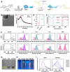
Synthesis and characterization of BiCD and BiCD-Ber. (A) Scheme of BiCD synthesis and the following conjugation with Ber-NH2 for BiCD-Ber. Part of the scheme was created in BioRender. (B) Transmission electron microscopy (TEM) image of BiCD with a lattice spacing of 0.31 nm. (C) X-ray diffraction (XRD) patterns of BiCD and BiCD-Ber with a wide angle (002) at approximately 25°. (D) ζ-potentials of BiCD and BiCD-Ber. (E) Fourier transform infrared (FTIR) spectra of BiCD, Ber-NH2, and BiCD-Ber. (F) X-ray photoelectron spectroscopy (XPS) analyses of BiCD and BiCD-Ber. Representative (G) optical and (H) fluorescent images of BiCD, Ber-NH2, and BiCD-Ber. (I) Fluorescent spectra of BiCD and BiCD-Ber.
Figure 2
Figure 2
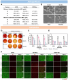
Antimicrobial and antibiofilm effects of BiCD, Ber-NH2, and BiCD-Ber. (A) Minimal inhibitory concentrations (MICs, μg/mL) and minimum bactericidal concentrations (MBCs, μg/mL) of BiCD, Ber-NH2, and BiCD-Ber against planktonicP. gingivalis(Pg),A. actinomycetemcomitans(Aa), andF. nucleatum(Fn). The assay was performed on three different occasions in duplicate, and the data are presented as the mean ± standard deviation (SD). (B) Morphology of Pg with or without treatment of BiCD, Ber-NH2, and BiCD-Ber at sub-MIC concentrations was assessed using field emission scanning electron microscopes (FE-SEM). (D) The 3-day-old Pg biofilms were treated with BiCD, Ber-NH2, and BiCD-Ber at different concentrations for 24 h, representative bacterial spots from Pg biofilms with/without treatments under different dilution factors showed the efficacy of treatments and reduction of Pg cells, and (C) the live bacteria in the biofilms were presented as log10 of the colony-forming units (CFUs). The assay was performed on three different occasions in duplicate, and the data are presented as mean ± SD. The asterisk (*) indicates the significant differences between the treatment and control groups (p < 0.05). (E) The thickness of Pg biofilms was determined from (F) the confocal images of the 3-day-old biofilms with/without treatments (scale bar: 50 μm).
Figure 3
Figure 3
Clearance of extracellular and/or intracellular Pg and the accumulation of nanomedicines in host cells. (A) Illustration of the Pg infection, antibiotic washing, treatment duration, and evaluation in studying the clearance of extracellular and/or intracellular Pg. Part of the scheme was created in BioRender. (B) The number of live bacteria in Pg-infected (B) pHGF or (D) HGECs with (+M/G) or without (−M/G) antibiotics washing followed by treatments with BiCD, Ber-NH2, and BiCD-Ber for a certain period. The assay was conducted on three different occasions in triplicate, and the data are presented as mean ± SD. The asterisk (*) and pound (#) signs reflect the significant differences between the treatment and control groups with p-value <0.05 or 0.0001, respectively. Representative colony formation images of (C) pHGF or (E) HGECs lysis samples showing the eradication efficiency of the treatments. (F) Fluorescent images of BiCD, Ber-NH2, and BiCD-Ber-treated pHGF or HGECs for 4 h (scale bar: 50 μm). (G) Bismuth mass in pHGF or HGECs after being treated with BiCD or BiCD-Ber at 50 μg/mL for 2, 6, and 24 h. The experiment was performed on three different occasions in triplicate, and the figure is generated from one repeat; data are presented as mean ± SD.
Figure 4
Figure 4
Bi-doped nanomedicines restored Pg-perturbed immuno-inflammatory response and protected against Pg-mediated disruption of cell attachment. (A) Schematic illustration of IL-1β-primed pHGF infected by Pg with/without treatments, followed by ELISA analysis of the pro-inflammatory cytokine levels in the supernatants; and the (B) concentrations of IL-8 and IL-6 in the supernatants from one representative biological repeat displayed as mean ± SD. The treatments and analysis were conducted for three different occasions in triplicate. (C) The effect of Ber-NH2, BiCD and BiCD-Ber on Pg-mediated HGEC detachment was investigated as illustrated. (D) The optical images of the cells infected by Pg with or without treatments indicate the cell attachment and junction status (scale bar: 200 μm). (E) Immunofluorescent staining of integrin β1 in HGECs followed by the previously mentioned treatments (scale bar: 50 μm). (F) Representative Western blots of integrin β1, E-cadherin, and GAPDH further revealed the effects of treatments on protecting against Pg-mediated digestion of cell-surface ligands. (G) The intensity of different protein blots was calculated from three biological repeats and statistically analyzed with the data presented as mean ± SD. The asterisk (*) indicates the significant differences between the control/treatment groups with the Pg group with p-value <0.05. Part of the scheme was created in BioRender.
Figure 5
Figure 5
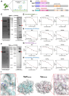
(A) Pg could generate outer membrane vesicles containing different membrane proteins and various virulence factors including gingipains. Part of the scheme was created in BioRender. (B) Schematic diagram of the structural domains of different gingipains. (C) Well-expressed and purified RgpB230–736 was examined by using Coomassie brilliant blue (CBB) staining and Western blot analysis. Red arrows pointing at the protein bands with their correlated molecular weight. The inhibitory effects of BiCD, Ber-NH2, and BiCD-Ber were investigated on (D) the amidolytic activity of recombinant RgpB230–736 and (E) the arginine-specific amidolytic activity of Pg suspension. (F) Recombinant Kgp229–595 was also expressed and examined using CBB and Western blot analysis. The suppression by BiCD, Ber-NH2, and BiCD-Ber on (G) the recombinant Kgp229–595 and (H) the lysine-specific amidolytic activity of the Pg suspension was studied in vitro. (I) Crystal structures of RgpB230–664 (PDB:1CVR) and Kgp229–680 (PDB:4TKX) with their catalytic pockets (red dashed square) showing the active sites composed of a histidine and a cysteine residue.
Figure 6
Figure 6
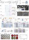
Construction of injectable Pg-responsive hydrogel for delivering BiCD-Ber. (A) A two-step structure modification of hyaluronic acid (HA) was conducted to obtain a disulfide-modified and acrylated HA derivative (HASSAC). (B) The successful modifications were confirmed by using 1H-NMR. (C) The proposed HA hydrogel was synthesized by photo-cross-linking HASSAC, and part of the scheme was created in BioRender. (D) The aqueous solution of HASSAC lost its fluidity after blue-light irradiation, while the hydrogel prepared from HASSAC at 1.5% (w/v) was injectable. (E) The injectable hydrogel with encapsulation of BiCD-Ber exhibited excellent shape adaptability and writing capability, as well as fluorescence under excitation. (F) SEM images of fabricated hydrogel displayed its porous structure, enabling the encapsulation of as-synthesized nanomedicines. (G) The constructed hydrogel loaded with red dye was proposed to degrade by disulfide-reducing reagents, such as TCEP and DTT, which were verified in subsequent experiments. (H) Pg was able to produce reductive sulfide metabolites and degrade hydrogel (upper scheme), and Pg-induced hydrogel degradation was compared to theS. mutans(Sm)-cocultured hydrogel for 6 days to verify the pathogen-specific degradation mode. (I) The BiCD-Ber encapsulated HA gel was designed to release BiCD-Ber to eliminate Pg while keeping the hydrogel intact (upper scheme), as confirmed in a 6-day incubation with Pg suspension. (J) BiCD-Ber HA gel with varying BiCD-Ber concentrations was tested against Pg at 108 CFU/mL for 3 days. (K) After treatment, the supernatants were collected and applied onto blood agar plates followed by a 7-day anaerobic culture for determining the anti-Pg effects of BiCD-Ber HA gel. (L) Releasing profiles of BiCD-Ber from BiCD-Ber HA gel (2 mg/mL) with or without TCEP as a disulfide-reducing reagent.
Figure 7
Figure 7
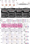
Therapeutic effects of BiCD-Ber HA gel on experimental periodontitis in vivo. (A) Schematic illustrations of the grouping and correspondent treatments in each group. (B) Experimental schedule of the in vivo study. Part of the scheme was created in BioRender. (C) Representative reconstructed microcomputed tomography (micro-CT) and sagittal images of the maxillary molars from each group (scale bar: 1 mm). Cementum-enamel junction (CEJ) and alveolar bone crest (ABC) were highlighted with red dashed lines. (D) The distance between the CEJ and ABC (CEJ-ABC), bone volume per total volume (BV/TV), trabecular separation (Tb. Sp), trabecular thickness (Tb. Th), and trabecular number (Tb. N) of maxillary alveolar bone surrounding the second molar of each group. (E) Representative histological staining images displaying periodontal tissue of maxillary second molars from different groups, with lines indicating the distance between CEJ and ABC (scale bar: 500 μm). (F) Statistical analysis of the distance between CEJ and ABC obtained from histological staining (n = 10 per group). (G) Representative TRAP staining images of each group. Black arrows indicate the positively stained multinucleated osteoclasts (scale bars: 100 μm). (H) Statistical analysis of the active osteoclast numbers surrounding the second molar of each group (n = 10 per group). (I) Representative immunohistochemical staining of IL-6, IL-1β, and IL-10 at gingiva tissues from each group (scale bar: 200 μm) and corresponding statistical analysis of each cytokine (n = 60 per group). The immunohistochemical images were analyzed using ImageJ. For each sample, six regions of interest (ROIs) were randomly selected, and color deconvolution was applied to isolate 3,3′-diaminobenzidine (DAB) staining. The positive area percentage was calculated using thresholding, ensuring consistent parameters across all images. All the data in the bar charts are presented as mean ± SD, and the statistical analyses are presented as asterisk (*) and pound (#) signs with p-value <0.05 or 0.0001, respectively.
Figure 8
Figure 8
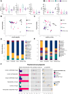
(A) Alpha diversity metrics of Shannon, Simpson, Ace, Chao, and Sobs among different groups. Principal coordinates analysis (PCoA) of (B) the weighted UniFrac and (C) unweighted UniFrac distance categorized by different groups. The relative abundance of periodontal microbiota at the (D) phylum and (E) genus levels among different groups. The phyla/genera with a relative abundance of less than 0.5% in the samples are merged into the ″Others″ item. (F) The relative abundance of Pg among different groups and the relevant analyses. The left bar chart shows the comparison between two randomly assigned groups, and the middle column displays the log2 value of the average relative abundance ratio between every two groups, while the right column presents the p and FDR values of the comparison.

References

    1. GBD 2021 Oral Disorders Collaborators. Trends in the Global, Regional, and National Burden of Oral Conditions from 1990 to 2021: A Systematic Analysis for the Global Burden of Disease Study 2021 Lancet 2025,405( (10482), ), 897-910. 10.1016/S0140-6736(24)02811-3. - DOI - PubMed
    1. Jin L.; Lamster I.; Greenspan J.; Pitts N.; Scully C.; Warnakulasuriya S. Global Burden of Oral Diseases: Emerging Concepts, Management and Interplay with Systemic Health. Oral Dis. 2016, 22 (7), 609–619. 10.1111/odi.12428. - DOI - PubMed
    1. Wolf T. G.; Cagetti M. G.; Fisher J.-M.; Seeberger G. K.; Campus G. Non-communicable Diseases and Oral Health: An Overview. Front. Oral. Health. 2021, 2, 725460.10.3389/froh.2021.725460. - DOI - PMC - PubMed
    1. Orlandi M.; Muñoz Aguilera E.; Marletta D.; Petrie A.; Suvan J.; D’Aiuto F. Impact of the Treatment of Periodontitis on Systemic Health and Quality of Life: A Systematic Review. J. Clin. Periodontol. 2021, 49 (S24), 314–327. 10.1111/jcpe.13554. - DOI - PubMed
    1. Thakkar-Samtani M.; Heaton L. J.; Kelly A. L.; Tayloar S. D.; Vidone L.; Tranby E. P. Periodontal treatment associated with decreased diabetes mellitus–related treatment costs. J. Am. Dent. Assoc. 2023, 154 (4), 283–292.e1. 10.1016/j.adaj.2022.12.011. - DOI - PubMed

Publication types