Assessing the effectiveness of texture and color enhancement imaging versus white-light endoscopy in detecting gastrointestinal lesions: A systematic review and meta-analysis
- PMID: 40313348
- PMCID: PMC12044138
- DOI: 10.1002/deo2.70128
Assessing the effectiveness of texture and color enhancement imaging versus white-light endoscopy in detecting gastrointestinal lesions: A systematic review and meta-analysis
Abstract
Introduction: Gastrointestinal cancers account for 26% of cancer incidence and 35% of cancer-related deaths globally. Early detection is crucial but often limited by white light endoscopy (WLE), which misses subtle lesions. Texture and color enhancement imaging (TXI), introduced in 2020, enhances texture, brightness, and color, addressing WLE's limitations. This meta-analysis evaluates TXI's effectiveness compared to WLE in gastrointestinal lesion lesion detection.
Methods: A systematic review and meta-analysis were conducted per Preferred Reporting Items for Systematic Reviews and Meta-Analyses guidelines. Searches of CENTRAL, PubMed, Embase, and Web of Science identified randomized controlled trials and observational studies comparing TXI with WLE. Outcomes included lesion detection rates, color differentiation, and visibility scores. The risk of bias was assessed using the Cochrane ROB 2.0 tool and Newcastle-Ottawa tools, and evidence certainty was evaluated using Grading of Recommendations Assessment, Development, and Evaluation.
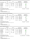
Results: Seventeen studies with 16,634 participants were included. TXI significantly improved color differentiation (mean difference: 3.31, 95% confidence interval [CI]: 2.49-4.13), visibility scores (mean difference: 0.50, 95% CI: 0.36-0.64), and lesion detection rates (odds ratio [OR]: 1.84, 95% CI: 1.52-2.22) compared to WLE. Subgroup analyses confirmed TXI's advantages across pharyngeal, esophageal, gastric, and colorectal lesions. TXI also enhanced adenoma detection rates (OR: 1.66, 95% CI: 1.31-2.12) and mean adenoma detection per procedure (mean difference: 0.48, 95% CI: 0.25-0.70).
Conclusion: TXI improves gastriontestinal lesion lesion detection by enhancing visualization and color differentiation, addressing key limitations of WLE. These findings support its integration into routine endoscopy, with further research needed to compare TXI with other modalities and explore its potential in real-time lesion detection.
Keywords: diagnostic imaging; early detection of cancer; gastrointestinal endoscopy; gastrointestinal neoplasms; image enhancement.
© 2025 The Author(s). DEN Open published by John Wiley & Sons Australia, Ltd on behalf of Japan Gastroenterological Endoscopy Society.
Conflict of interest statement
None.
Figures


References
-
- Aalami AH, Abdeahad H, Mesgari M. Circulating miR‐21 as a potential biomarker in human digestive system carcinoma: A systematic review and diagnostic meta‐analysis. Biomarkers 2021; 26: 103–13. Available from: https://www.tandfonline.com/doi/full/10.1080/1354750X.2021.1875504 - DOI - PubMed
-
- Choi HS, Hwang JH. Endoscopic resection of early luminal cancer. Gastrointest Endosc Clin N Am 2024; 34: 51–78. Available from: https://linkinghub.elsevier.com/retrieve/pii/S1052515723000661 - PubMed
-
- Akarsu M, Akarsu C. Evaluation of new technologies in gastrointestinal endoscopy. J Soc Laparoendosc Surg 2018; 22 :e2017.00053. Available from: https://www.ncbi.nlm.nih.gov/pmc/articles/PMC5788542/ - PMC - PubMed
-
- Dobashi A, Ono S, Furuhashi H et al. Texture and Color enhancement imaging increases color changes and improves visibility for squamous cell carcinoma suspicious lesions in the pharynx and esophagus. Diagnostics 2021; 11: 1971. Available from: https://www.mdpi.com/2075‐4418/11/11/1971 - PMC - PubMed
-
- Antonelli G, Bevivino G, Pecere S et al. Texture and color enhancement imaging versus high definition white‐light endoscopy for detection of colorectal neoplasia: A randomized trial. Endoscopy 2023; 55: 1072–80. Available from: https://pubmed.ncbi.nlm.nih.gov/37451283/ - PubMed
Publication types
LinkOut - more resources
Full Text Sources
