Review
. 2025 Apr 23:2025:5871029.
doi: 10.1155/crp/5871029. eCollection 2025.

Extracardiac Compression by Gastrointestinal Structures: A Comprehensive Anthology From the Literature

Affiliations
Review

Extracardiac Compression by Gastrointestinal Structures: A Comprehensive Anthology From the Literature

Riccardo Scagliola et al. Cardiol Res Pract. .

Abstract

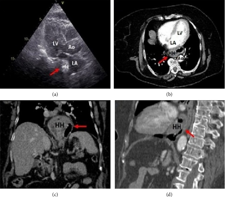
Extrinsic heart compression by gastrointestinal (GI) structures is an often underrecognized finding in clinical practice. It is potentially related to unpredictable clinical conditions, ranging from incidental detection in asymptomatic subjects, to deranging and potentially life-threatening clinical manifestations. However, despite its potential clinical relevance, there is still no comprehensive analysis investigating the surrounding causes, clinical findings, and diagnostic imaging work-up for this patient population. A narrative review with an extensive bibliographic search of the literature was performed using PubMed (MEDLINE), Embase, and Cochrane Central Databases up to December 31, 2023. Despite the broad spectrum of GI etiologies, clinical manifestations, and cardiac chamber involvement scenarios, physicians must be aware of such an uncommon condition, in order to provide timely diagnosis through a comprehensive imaging approach, avoid misleading interpretations, and determine the most appropriate decision-making strategy.

Keywords: ECG changes; clinical findings; extracardiac compression; gastrointestinal structures; imaging tools.


Conflict of interest statement

The authors declare no conflicts of interest.

Figures

Figure 1
Figure 1
(a) Transthoracic echocardiography, parasternal long-axis view showing an echolucent extrinsic mass compressing the posterior aspect of the left atrial wall (red arrow). (b–d) Thoracoabdominal computed tomography showing extrinsic left atrial compression by a large hiatal hernia, respectively, at the transverse, coronal, and sagittal sections through the left atrium (red arrow). Abbreviations: Ao: aorta; HH: hiatal hernia; LA: left atrium; LV: left ventricle.
Figure 2
Figure 2
(a) Chest X-ray showing a convex opacity overlapping the right mediastinum (red oval) and the absence of a gastric bubble (green oval). (b) Chest computed tomography, sagittal section through the left atrium showing tracheal and left atrial compression from an enlarged esophagus (blue oval). (c) Barium swallowing radiogram showing a “bird's beak” or “rat's tail” appearance of the lower esophagus (yellow circle) with no contrast trickling into the stomach (adapted from Bhamrah et al.) [10].
Figure 3
Figure 3
(a) Barium swallowing radiogram showing proximal esophageal dilation (red arrow) and midesophageal narrowing (blue arrow). (b) Chest computed tomography, sagittal section through the left atrium showing significant left atrial compression from an esophageal mass (red arrow) (adapted from Polina et al.) [12]. Abbreviations: LA: left atrium; LV: left ventricle.
Figure 4
Figure 4
(a) Barium swallowing radiogram showing laparoscopic adjustable gastric banding rotation resulting in band slippage (red circle). (b) Transthoracic echocardiography, apical 4-chamber view showing left atrial compression from megaesophagus during diastole (red asterisk). (c) Computed tomography, transverse section showing megaesophagus causing extrinsic left atrial compression (red arrow) (adapted from De Silva et al.) [19].
Figure 5
Figure 5
(a) Fluoroscopy showing the radiologic appearance of a stent-in-stent at the time of esophageal stent implantation. (b and c) Computed tomography, transverse, and sagittal sections showing partial compression of the posterior left atrial wall by an esophageal stent. Note the termination of the inner stent at left atrium level (red arrow) (adapted from Mazzella et al.) [20].
Figure 6
Figure 6
(a) Computed tomography, coronal section showing right diaphragmatic herniation (blue arrow). (b) Computed tomography, transverse section showing digestive handles in the right thorax cavity compressing the right-side heart chambers (blue arrow) (adapted from Deniau and Beloucif) [42].
Figure 7
Figure 7
(a) Transthoracic echocardiography, apical 4-chamber view showing a mass of uncertain origin (intra-atrial vs. extracardiac) at left atrial posterior wall level (large arrows). (b) The same echocardiographic projection immediately after ingestion of a carbonated beverage showing the mass entirely filled with bubbles, thus confirming its extracardiac location (small arrows) (adapted from Bouzas-Mosquera et al.) [74]. Abbreviations: LV: left ventricle; RA: right atrium; RV: right ventricle.
Figure 8
Figure 8
Flowchart summing up a proposed decision-making approach in this patient population.

References

    1. D’Cruz I. A., Feghali N., Gross C. M. Echocardiographic Manifestations of Mediastinal Masses Compressing or Encroaching on the Heart. Echocardiography . 1994 September;11(5):523–533. doi: 10.1111/j.1540-8175.1994.tb01093.x. - DOI - PubMed
    1. van Rooijen J. M., van den Merkhof L. F. Left Atrial Impression: A Sign of Extra-Cardiac Pathology. European Journal of Echocardiography . 2008 September;9(5):661–664. doi: 10.1093/ejechocard/jen031. - DOI - PubMed
    1. Raza S. T., Mukherjee S. K., Danias P. G., et al. Hemodynamically Significant Extrinsic Left Atrial Compression by Gastric Structures in the Mediastinum. Annals of Internal Medicine . 1995 July;123(2):114–116. doi: 10.7326/0003-4819-123-2-199507150-00006. - DOI - PubMed
    1. Mitiek M. O., Andrade R. S. Giant Hiatal Hernia. The Annals of Thoracic Surgery . 2010 June;89(6):S2168–S2173. doi: 10.1016/j.athoracsur.2010.03.022. - DOI - PubMed
    1. Skinner D. B., Belsey R. H., Russell P. S. Surgical Management of Esophageal Reflux and Hiatus Hernia. The Journal of Thoracic and Cardiovascular Surgery . 1967 January;53(1):33–54. doi: 10.1016/s0022-5223(19)43239-x. - DOI - PubMed

LinkOut - more resources