Review
. 2025 Apr 17:13:1577081.
doi: 10.3389/fcell.2025.1577081. eCollection 2025.

Impact of mitochondrial metabolism on T-cell dysfunction in chronic lymphocytic leukemia

Affiliations
Review

Impact of mitochondrial metabolism on T-cell dysfunction in chronic lymphocytic leukemia

Wael Gamal et al. Front Cell Dev Biol. .

Abstract

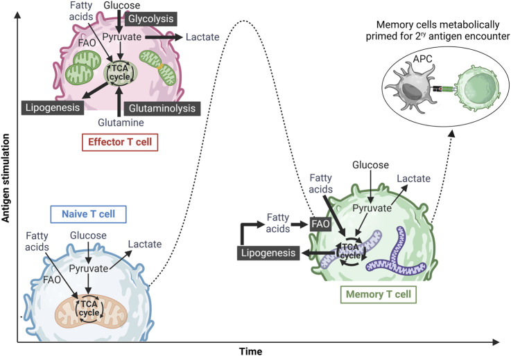
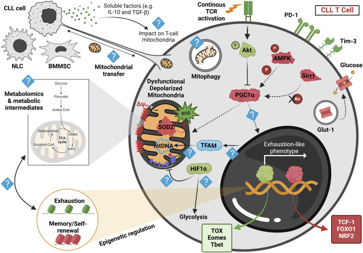
T cells play a central role in anti-tumor immunity, yet their function is often compromised within the immunosuppressive tumor microenvironment, leading to cancer progression and resistance to immunotherapies. T-cell activation and differentiation require dynamic metabolic shifts, with mitochondrial metabolism playing a crucial role in sustaining their function. Research in cancer immunometabolism has revealed key mitochondrial abnormalities in tumor-infiltrating lymphocytes, including reduced mitochondrial capacity, depolarization, structural defects, and elevated reactive oxygen species. While these mitochondrial disruptions are well-characterized in solid tumors and linked to T-cell exhaustion, their impact on T-cell immunity in lymphoproliferative disorders remains underexplored. Chronic lymphocytic leukemia (CLL), the most prevalent chronic adult leukemia, is marked by profound T-cell dysfunction that limits the success of adoptive cell therapies. Emerging studies are shedding light on the role of mitochondrial disturbances in CLL-related T-cell dysfunction, but significant knowledge gaps remain. This review explores mitochondrial metabolism in T-cell exhaustion, emphasizing recent findings in CLL. We also discuss therapeutic strategies to restore T-cell mitochondrial function and identify key research gaps.

Keywords: CAR T cell; CLL (chronic lymphocytic leukemia); T-cell exhaustion; adoptive cell immunotherapy; cancer; metabolism; mitochondria.


Conflict of interest statement

The authors declare that the research was conducted in the absence of any commercial or financial relationships that could be construed as a potential conflict of interest.

Figures

FIGURE 1
FIGURE 1
Metabolic reprogramming of T cells upon antigen stimulation. Naïve T cells exist in a metabolically quiescent state, primarily utilizing basal oxidative phosphorylation (OXPHOS) to meet their minimal energy demands. Upon antigen recognition and activation, these cells differentiate into effector T cells, undergoing a metabolic shift toward an anabolic state characterized by increased aerobic glycolysis, glutamine oxidation, and OXPHOS to support rapid expansion and effector functions. Effector T cells also exhibit mitochondrial fission, mediated by Drp-1, to accommodate heightened metabolic demands. Following antigen clearance and resolution of the immune response, a subset of surviving T cells differentiates into memory T cells, reverting to a metabolically quiescent state predominantly reliant on OXPHOS and fatty acid oxidation (FAO) for long-term survival and energy homeostasis. Memory T cells can also utilize glucose-derived ATP to synthesize fatty acids, which are subsequently broken down via FAO, a process essential for their long-term maintenance. Additionally, these cells are characterized by fused mitochondrial networks with high spare respiratory capacity (SRC), enabling them to rapidly respond to subsequent antigenic challenges. Figure was created in BioRender. Gamal et al. (2025), https://BioRender.com/p2qlxtp.
FIGURE 2
FIGURE 2
Proposed model of mitochondrial dysregulation in CLL-derived T cells at the basal state, highlighting current findings and future research directions. Human and murine CLL T cells accumulate depolarized mitochondria, leading to dysfunctional mitochondrial activity post in vitro activation. This depolarization correlates with elevated reactive oxygen species (ROS) levels, likely due to reduced expression of superoxide dismutase 2 (SOD2) observed in both human and murine cells. These mitochondrial disturbances, coupled with reduced levels of the transcription factor NRF2—an essential regulator of oxidative metabolism—are associated with the downregulation of PGC1α, the key regulator of mitochondrial function. Persistent stimulation of CLL T cells within the leukemic microenvironment is thought to activate Akt signaling, as recently demonstrated in resting CLL T cells from the adoptive transfer (AT) Eμ-TCL1 murine model, which may subsequently suppress PGC1α activity. Furthermore, recent findings suggest a downregulation of AMPK phosphorylation and lower Sirt1 expression in AT Eμ-TCL1 T cells, potentially impairing PGC1α function by limiting its phosphorylation and deacetylation, respectively. These metabolic alterations are also associated with decreased intracellular Glut-1 reserves in human CLL T cells, potentially hindering glucose uptake upon in vitro activation. Such metabolic disturbances correlate with an exhaustion-like T-cell differentiation phenotype, marked by increased expression of TOX, Eomes, and T-bet transcription factors, along with elevated PD-1 and Tim-3 levels, while displaying a diminished memory and stemness profile, as indicated by reduced expression of TCF-1 and increased FOXO1 phosphorylation. Despite these insights, key aspects of mitochondrial dysfunction in CLL T cells remain unclear. For instance, the influence of soluble factors such as IL-10 and TGF-β, released by CLL cells, on T-cell mitochondrial activity is unknown. Furthermore, the precise role of PGC1α in driving T-cell exhaustion remains to be investigated. Additionally, TFAM, a crucial mitochondrial transcription factor, warrants further study, particularly given recent findings that suggest reduced chromatin accessibility near TFAM in AT Eμ-TCL1 T cells. Studies in human CLL patient-derived T cells have indicated increased HIF1α expression; however, it remains to be determined whether this upregulation of hypoxic signature contributes to suppressed OXPHOS and enhanced glycolysis, as observed in solid tumors. Moreover, metabolomic analyses are needed to identify mitochondrial metabolite dysregulation in CLL-derived T cells and its relationship with the epigenetic changes driving exhaustion. Finally, exploring nanotube formation and mitochondrial transfer between T cells and microenvironmental cells may provide novel insights into CLL-associated metabolic dysregulation. Red represents downregulated proteins, green signifies upregulated proteins, and light blue indicates an unknown mechanism. Dashed arrows denote reduced regulation. NLC, nurse-like cell; BMMSC, bone marrow mesenchymal stromal cell. Figure was created in BioRender. Gamal et al. (2025), https://BioRender.com/lkbyyip.

Similar articles

References

    1. Abdel-Hakeem M. S., Manne S., Beltra J.-C., Stelekati E., Chen Z., Nzingha K., et al. (2021). Epigenetic scarring of exhausted T cells hinders memory differentiation upon eliminating chronic antigenic stimulation. Nat. Immunol. 22 (8), 1008–1019. 10.1038/s41590-021-00975-5 - DOI - PMC - PubMed
    1. Ahearne M. J., Willimott S., Piñon L., Kennedy D. B., Miall F., Dyer M. J., et al. (2013). Enhancement of CD 154/IL 4 proliferation by the T follicular helper (T fh) cytokine, IL 21 and increased numbers of circulating cells resembling T fh cells in chronic lymphocytic leukaemia. Br. J. Haematol. 162 (3), 360–370. 10.1111/bjh.12401 - DOI - PubMed
    1. Al-Awar A., Hussain S. (2024). Interplay of reactive oxygen species (ROS) and epigenetic Remodelling in Cardiovascular diseases pathogenesis: a Contemporary perspective. FBL 29 (11), 398. 10.31083/j.fbl2911398 - DOI - PubMed
    1. Baitsch L., Baumgaertner P., Devêvre E., Raghav S. K., Legat A., Barba L., et al. (2011). Exhaustion of tumor-specific CD8⁺ T cells in metastases from melanoma patients. J. Clin. Investigation 121 (6), 2350–2360. 10.1172/JCI46102 - DOI - PMC - PubMed
    1. Baixauli F., Martín-Cófreces N. B., Morlino G., Carrasco Y. R., Calabia-Linares C., Veiga E., et al. (2011). The mitochondrial fission factor dynamin-related protein 1 modulates T-cell receptor signalling at the immune synapse. Embo J. 30 (7), 1238–1250. 10.1038/emboj.2011.25 - DOI - PMC - PubMed

LinkOut - more resources