Review
. 2025 Sep;57(5):1183-1203.
doi: 10.1111/evj.14508. Epub 2025 May 2.

Magnetic resonance imaging for diagnosing and managing deep digital flexor tendinopathy in equine athletes: Insights, advances and future directions

Affiliations
Review

Magnetic resonance imaging for diagnosing and managing deep digital flexor tendinopathy in equine athletes: Insights, advances and future directions

Alexandra Scharf et al. Equine Vet J. 2025 Sep.

Abstract

Deep digital flexor (DDF) tendinopathy is a significant cause of lameness and poor performance in equine athletes with substantial implications for their return to athletic performance. Magnetic resonance imaging (MRI) is increasingly integrated into the diagnostic workup of horses with foot pain and has revolutionised the diagnosis and management of these injuries. This review discusses the principles of MRI in the context of deep digital flexor tendon (DDFT) injury, comparing high-field and low-field systems and highlighting the clinical relevance of technical parameters, including field strength and sequence selection, in achieving an accurate diagnosis. This review also critically evaluates how different configurations and/or imaging features of tendon lesions may impact patient prognosis, considers the complementary role of computed tomography and ultrasonography in cases where MRI may not be feasible, and discusses emerging imaging techniques including positron emission tomography (PET)-MRI and quantitative MRI. Lastly, this review underscores the importance of serial imaging to monitor lesion progression and guide rehabilitation, while identifying knowledge gaps and proposing future research directions. Ultimately, a multidisciplinary approach incorporating advanced imaging and tailored rehabilitation is essential to improving clinical outcomes in horses with DDFT injuries.

Keywords: MRI; horse; tendinopathy; tendon.


Conflict of interest statement

The authors declare no conflicts of interest.

Figures
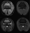
FIGURE 1
FIGURE 1
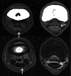
High‐field MRI (3.0 T) images of a 5‐year‐old Warmblood that initially presented for an MRI after a grade 2/5 left forelimb lameness developed at a competition 2 months prior. The lameness localised to palmar digital analgesia and did not respond to rest and injection of the distal interphalangeal joint. This horse was diagnosed with mild dorsal fibrillation of the lateral lobe of the deep digital flexor tendon (A, white arrow). The navicular bursa was injected with corticosteroids and an intravenous regional limb perfusion of mesenchymal stem cells was performed. The horse experienced complete resolution of lameness and returned to full work. Three years later, this horse returned to be evaluated for a more severe episode of left forelimb lameness. At this time, a large, axial tear with complete dorsal border disruption was seen in the lateral lobe just proximal to the original site of fibrillation (B, white arrowhead). There was also marked chronic navicular bursitis with probable adhesion formation. MRI sequences shown include a transverse proton density‐weighted in‐phase DIXON (left) and T1‐weighted volume interpolate breath hold (VIBE) (right).
FIGURE 2
FIGURE 2
Marked deep digital flexor tendinopathy. Transverse T1‐weighted gradient echo (A), proton density‐weighted (B), T2‐weighted (C) and short tau inversion recovery (STIR) (D) sequences acquired with a standing low‐field MRI are shown at the level of the collateral sesamoidean ligament. There is a large dorsal border tear in the lateral lobe and a thin parasagittal split in the medial lobe. The lateral lesion occupies nearly one‐half of the tendon lobe, has marked associated fluid signal (white arrow), and has an associated dorsal flap. The dorsal‐to‐palmar extent of the smaller, medial parasagittal split (arrowhead) is only seen in the T1‐weighted gradient echo sequence (A).
FIGURE 3
FIGURE 3
High‐field MRI (1.0 T) sagittal fast spin echo short tau inversion recovery (STIR) (A) and transverse fast spin echo proton density‐weighted (B) images highlighting palmar herniation of the navicular bursa through a parasagittal split of the deep digital flexor tendon with complete disruption of the dorsal and palmar borders (arrows).
FIGURE 4
FIGURE 4
High‐field MRI (3.0 T) short tau inversion recovery (STIR) (A) and proton density‐weighted in‐phase DIXON (B) images demonstrating grade 3 adhesion formation between the collateral sesamoidean ligament (open white arrow) and deep digital flexor tendon. The adhesions span the navicular bursa, which is not well‐defined. A large core lesion with associated dorsal border disruption is seen in the deep digital flexor tendon (solid white arrowhead), which is diffusely enlarged. Adhesions were confirmed at post‐mortem exam.
FIGURE 5
FIGURE 5
Fast spin echo proton density (PD) (A, C) and T2‐weighted (B, D) standing low‐field MRI images of a grand prix jumper obtained with a standing low‐field system. This horse presented 5 days after acute onset of lameness. MRI was performed at the time of presentation (A, B) and was repeated 25 days after the initial scan (C, D). At presentation, small regions of fibre disruption were identified in the lateral lobe of the DDFT at the level of the pastern (not shown), proximal recess of the navicular bursa (A) and insertion (not shown). At recheck, there was propagation of the tear with progressive fibre disruption in the lateral lobe despite appropriate rest and medical management.
FIGURE 6
FIGURE 6
Two comparative examples of deep digital flexor tendinopathy using fan beam computed tomography (A, B) and high‐field MRI at 1.5 T (C, D) at the level of the proximal phalanx. Proton density‐weighted transverse MR images are shown. Images A and C demonstrate a palmar sagittal split with palmar border disruption. Images B and D demonstrate a dorsal marginal tear at the junction of the ovoid and bilobed tendon.

Similar articles

References

    1. Ely ER, Avella CS, Price JS, Smith RKW, Wood JLN, Verheyen KLP. Descriptive epidemiology of fracture, tendon and suspensory ligament injuries in National Hunt racehorses in training. Equine Vet J. 2009;41:372–378. - PubMed
    1. Parkin TDH, Rogers K, Stirk A, et al. Risk factors for tendon strain injury during racing in Great Britain. Proceedings of the 16th International Conference of Racing Analysts and Veterinarians. Newmarket, UK: R and W Publications; 2006. p. 104.
    1. Williams RB, Harkins LS, Hammond CJ, Wood JL. Racehorse injuries, clinical problems and fatalities recorded on British racecourses from flat racing and National Hunt racing during 1996, 1997 and 1998. Equine Vet J. 2001;33:478–486. - PubMed
    1. Gutierrez‐Nibeyro SD, Werpy NM, Gold SJ, Olguin S, Schaeffer DJ. Standing MRI lesions of the distal interphalangeal joint and podotrochlear apparatus occur with a high frequency in warmblood horses. Vet Radiol Ultrasound. 2020;61:336–345. - PubMed
    1. Eliashar E, McGuigan MP, Wilson AM. Relationship of foot conformation and force applied to the navicular bone of sound horses at the trot. Equine Vet J. 2004;36:431–435. - PubMed

MeSH terms