. 2025 May 2;149(1):41.
doi: 10.1007/s00401-025-02871-w.

Abundant non-inclusion α-synuclein pathology in Lewy body-negative LRRK2-mutant cases

Affiliations

Abundant non-inclusion α-synuclein pathology in Lewy body-negative LRRK2-mutant cases

Nanna Møller Jensen et al. Acta Neuropathol. .

Abstract

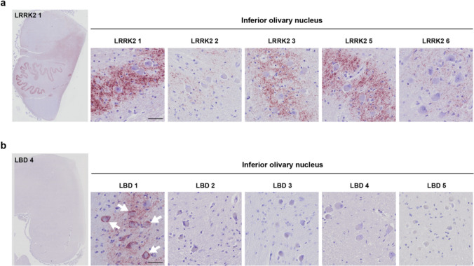
Lewy body diseases are common neurodegenerative diseases, including Parkinson's disease (PD) and dementia with Lewy bodies, which lead to both motor and non-motor symptoms. They are neuropathologically characterized by loss of neuromelanized neurons in the substantia nigra pars compacta and α-synuclein-immunopositive inclusions (Lewy bodies) in several types of neurons in the brain. A fraction of monogenic PD cases, however, represent a conundrum, as they can present with clinical Lewy body disease but do not have Lewy bodies upon neuropathological examination. For LRRK2, the presence or absence of Lewy bodies is not related to any specific mutation in the gene and different clinical presentation and neuropathology can be present even in the same family. Here, we present the first evidence of widespread α-synuclein accumulation detected with proximity ligation assay (PLA) using the MJFR14-6-4-2 antibody in six Lewy body-negative LRRK2 cases and compare the levels with five patients with neuropathologically verified Lewy body disease and six healthy controls. We show that non-inclusion aggregated α-synuclein in the form of particulate PLA signal is dominant in the LRRK2 cases, while both Lewy-like and particulate PLA signal is found in late-stage Lewy body disease. Furthermore, LRRK2 cases displayed prominent particulate PLA signal in pontocerebellar tracts and inferior olivary nuclei in the brainstem, which was not seen in idiopathic Lewy body disease cases. These results suggest that Lewy-body negative LRRK2-related PD is not associated with a lack of α-synuclein aggregation in neurons but rather a deficiency in the formation of inclusions.

Keywords: LRRK2; Lewy body disease; Neurodegeneration; Non-inclusion pathology; Proximity ligation assay; α-Synuclein.


Conflict of interest statement

Declarations. Conflict of interest: The authors declare no competing interests. Ethical approval and informed consent: Post-mortem brain tissue was obtained from Parkinson’s UK Brain Bank (PUKBB), Imperial College London, and Oxford Brain Bank (OBB), Nuffield Department of Clinical Neurosciences in University of Oxford, in accordance with approved protocols by the Wales Research Ethics Committee (23/WA/0273) and the Ethics Committee of the University of Oxford (ref 23/SC/0241). All participants had given prior written informed consent for the brain donation. Both brain banks comply with the requirements of the Human Tissue Act 2004 and the Codes of Practice set by the Human Tissue Authority (HTA licence numbers 12275 for PUKBB and 12217 for OBB).

Figures

Fig. 1
Fig. 1
Regions of interest and PLA terminology. a Five brain regions (medulla, pons, midbrain, posterior hippocampus, and amygdala) were included for each case to allow comparative quantitative analyses of PLA signals. See Suppl. Figure 3 for delineation of the listed subregions in three of the cases included in this study. b The PLA staining yielded two distinct patterns of signal: the particulate PLA not associated with inclusions (top panels) with increasing signal density from left to right and a Lewy-like PLA staining (bottom panel) with strong morphological similarity to LBs and LNs as stained by standard IHC. Presumed LBs are indicated by arrowheads, while examples of putative LNs are highlighted with arrows. Scale bar = 20 µm (applies to all images). Particulate and Lewy-like PLA were analysed separately in all sections and reported as area covered (%)
Fig. 2
Fig. 2
Particulate PLA, Lewy-like PLA, and LB densities in LRRK2 LB-negative cases, regular LBD, and non-neurodegenerative controls. a Overview of PLA staining appearance in medulla (around DMV), pons (around LC), midbrain (around SNpc), posterior hippocampus (around CA2), and amygdaloid complex (in the amygdala). Scale bar = 50 µm (applies to all images). b–c Quantitative analysis performed in the entire tissue sections showing particulate PLA area coverage (%, b) and Lewy-like PLA area coverage (%, c) for the three groups. All graphs show mean ± SEM with data points for individual cases. Univariate analyses adjusting for age and sex followed by Bonferroni’s multiple comparison test. *p < 0.05, **p < 0.01, ***p < 0.001. CA2 cornu ammonis 2 of the hippocampus, DMV dorsal motor nucleus of the vagus, LC locus coeruleus, SNpc (substantia nigra pars compacta)
Fig. 3
Fig. 3
PLA signal in specific brain regions from selected cases. Specific brain regions were identified from the medulla (dorsal motor nucleus of the vagus, reticular formation, and inferior olivary nucleus), pons (locus coeruleus and raphe nucleus), midbrain (substantia nigra pars compacta, SNpc, and ventral tegmental area, VTA), posterior hippocampus (CA3, CA2, and entorhinal cortex) and amygdala. Three cases were displayed from each group, encompassing most of the variation in signal densities in each group. Scale bar = 10 µm (applies to all images). Note that LRRK2 4 was almost entirely depigmented and had substantial neuronal loss in several regions, incl. SNpc. See also semi-quantitative overview particulate versus Lewy-like PLA in all cases in Fig. 6
Fig. 4
Fig. 4
Prominent PLA signal in the inferior olivary nucleus of LRRK2 cases. a LRRK2 cases displayed particulate PLA signal in the inferior olivary nucleus, with signal predominantly present in the neuropil surrounding the neuronal cell bodies. Note that LRRK2 4, which contained no PLA signal in the medulla, was left out of the figure. b Only LBD 1 of the LBD cases displayed PLA signal in the inferior olivary nucleus. Note that in this case, signal is not confined to the neuropil but also found in the neuronal cell bodies. Scale bars = 50 µm. See also semi-quantitative overview particulate versus Lewy-like PLA in all cases in Fig. 6
Fig. 5
Fig. 5
PLA in pontine nuclei, transverse and longitudinal fibres of the basilar pons. a Particulate and Lewy-like PLA signal was assessed in six ROIs in the basilar pons of all cases as indicated in the overviews from one LRRK2 case (left) and one LBD case (right). Magnified panels show transverse fibres (outlined in blue) and longitudinal fibres (outlined in yellow), while in-between areas correspond to pontine nuclei. Scale bars = 200 µm. b LRRK2 cases displayed dense particulate PLA signal in the transverse (pontocerebellar) fibres and to some degree in the pontine nuclei. Note that LRRK2 4, which was negative in the pons, was left out of the figure. Scale bar = 20 µm (applies to all images). c LBD cases displayed much less signal in pontine nuclei and transverse fibres than the LRRK2 cases. One case (LBD 5) presented with some Lewy-like PLA signal in the longitudinal fibre bundles (arrows). Scale bar = 20 µm (applies to all images). See also semi-quantitative overview particulate versus Lewy-like PLA in all cases in Fig. 6 and quantifications in Suppl. Figure 4
Fig. 6
Fig. 6
Graphical summary of particulate and Lewy-like PLA across all cases and nuclei/regions examined. Based on the quantifications of area coverage, semi-quantitative scales were made for particulate PLA (a) and Lewy-like PLA (b). Note that the same colour in a and b denotes a 300-fold difference in area %. LBD cases stand out with a high density of Lewy-like PLA across the regions examined, while LRRK2 cases display the highest particulate PLA density. N/A, not available
Fig. 7
Fig. 7
Cell loss and extra-neuronal neuromelanin in the locus coeruleus of LBD cases. Locus coeruleus displayed varying degrees of loss of its noradrenergic, neuromelanin-containing neurons in the LBD cases, as illustrated with a progressive fall in the density of melanized neurons (left to right). Extra-neuronal neuromelanin was apparent in some cases (arrowheads in LBD 2 and 5), evincing neuronal loss. PLA signal density was also lower in LBD cases with extensive neuronal loss (arrows indicate examples of Lewy-like PLA). Scale bar = 50 µm

References

    1. Altay MF, Liu AKL, Holton JL, Parkkinen L, Lashuel HA (2022) Prominent astrocytic alpha-synuclein pathology with unique post-translational modification signatures unveiled across Lewy body disorders. Acta Neuropathol Commun 10:1–18. 10.1186/s40478-022-01468-8 - PMC - PubMed
    1. Amunts K, Kedo O, Kindler M, Pieperhoff P, Mohlberg H, Shah NJ et al (2005) Cytoarchitectonic mapping of the human amygdala, hippocampal region and entorhinal cortex: Intersubject variability and probability maps. Anat Embryol (Berl) 210:343–352. 10.1007/S00429-005-0025-5 - PubMed
    1. Bankhead P, Loughrey MB, Fernández JA, Dombrowski Y, McArt DG, Dunne PD et al (2017) QuPath: Open source software for digital pathology image analysis. Sci Rep 7:1–7. 10.1038/s41598-017-17204-5 - PMC - PubMed
    1. Beach TG, Adler CH, Lue LF, Sue LI, Bachalakuri J, Henry-Watson J et al (2009) Unified staging system for Lewy body disorders: Correlation with nigrostriatal degeneration, cognitive impairment and motor dysfunction. Acta Neuropathol 117:613–634. 10.1007/s00401-009-0538-8 - PMC - PubMed
    1. Bengoa-Vergniory N, Roberts RF, Wade-Martins R, Alegre-Abarrategui J (2017) Alpha-synuclein oligomers: a new hope. Acta Neuropathol 134:819–838. 10.1007/s00401-017-1755-1 - PMC - PubMed

Publication types

Substances