. 2025 May 2;11(18):eado6644.
doi: 10.1126/sciadv.ado6644. Epub 2025 May 2.

Development of the mechanoresponsive pericellular matrix of chondrons

Affiliations

Development of the mechanoresponsive pericellular matrix of chondrons

Donghee Lee et al. Sci Adv. .

Abstract

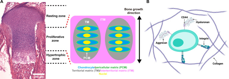
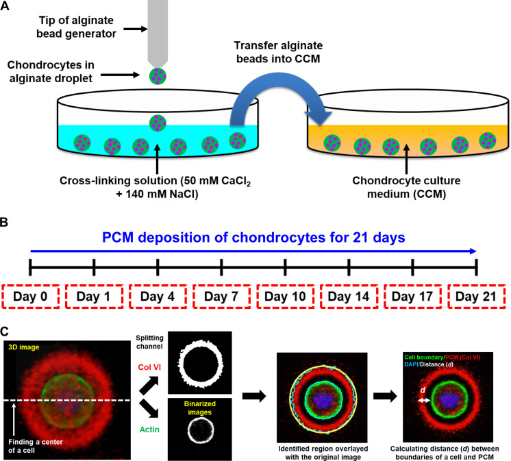
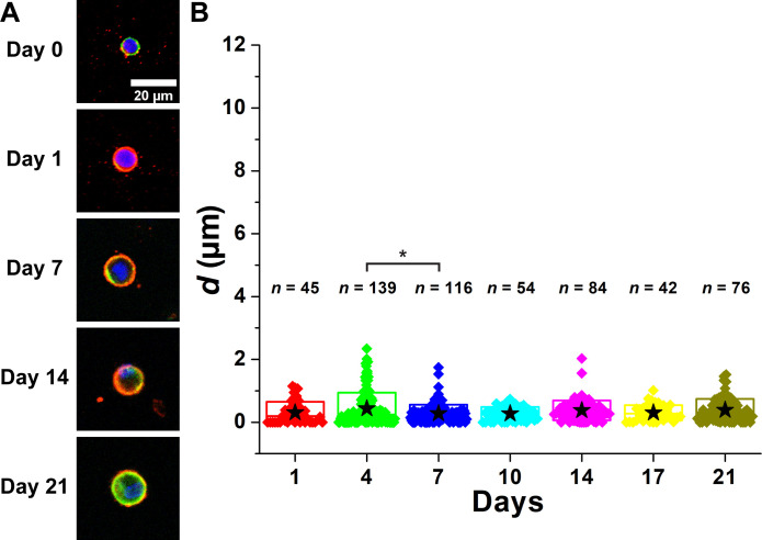
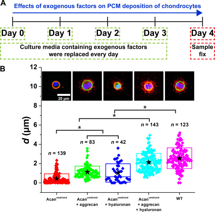
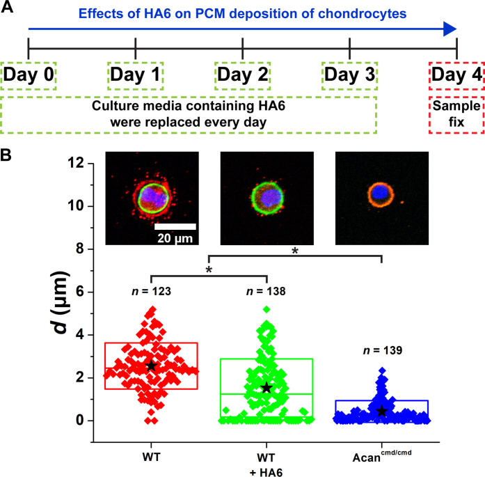
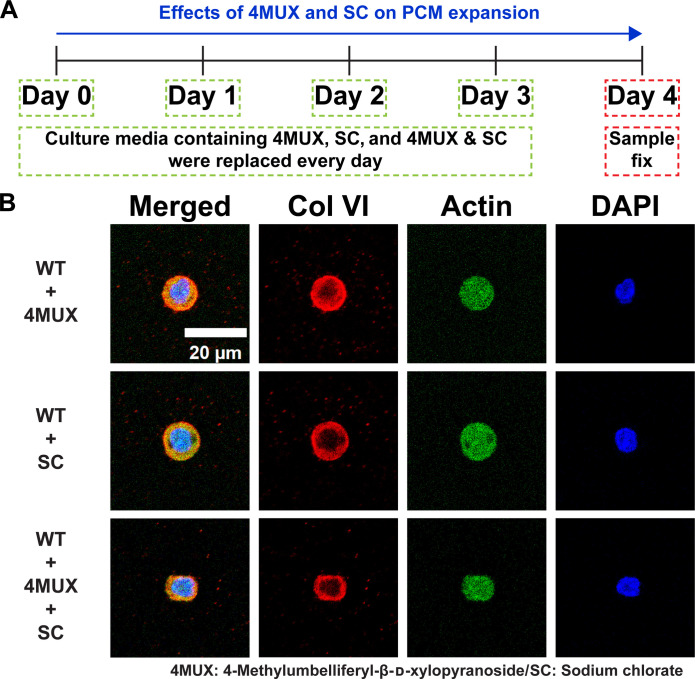
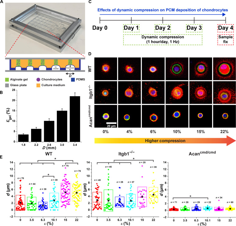
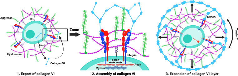
Physical properties of cartilage are conferred by the composition and ultrastructure of the extracellular matrix. This study focuses on the development of the pericellular matrix (PCM), a domain that directly contacts the chondrocyte and is a key regulator of biomechanical and biochemical signaling. Using three-dimensional cell culture, microfluidic cell compression platforms, and genetic mouse models, we demonstrated that collagen VI is initially assembled at the cell surface and then displaced to form a shell at the PCM-territorial matrix boundary. Cell surface-bound hyaluronan is crucial for the assembly process, and hyaluronan-aggrecan complexes drive displacement. Integrin adhesion is not required early but is crucial to determine the final placement of the collagen VI shell. Dynamic compression accelerated PCM maturation except in aggrecan mutants. Together, these findings provide key insights into the development of the mechanosensitive PCM and establish an in vitro platform to support studies of matrix biology in normal and disease models.


Figures

Fig. 1.
Fig. 1.. Matrix structure of growth plate cartilage.
(A) Hematoxylin and eosin staining of a growth plate cartilage in a hindlimb proximal tibia and schematic of matrix architecture of growth plate. Chondrocytes are encapsulated by PCM, and chondrocyte columns are surrounded by a territorial matrix (TM). The chondrocyte columns are separated by interterritorial matrix (ITM). (B) Schematic of matrix molecules around a chondrocyte. Collagens attach to the chondrocyte via integrin receptors. HA binds to the cell surface through the CD44 receptor. The CD44 receptor is a cell surface glycoprotein that serves as a receptor for HA, allowing HA to interact with the cell membrane. In addition, aggrecan proteins covalently bind to HA through a link protein.
Fig. 2.
Fig. 2.. Systems for studying PCM deposition of chondrocytes.
(A) Chondrocytes were 3D-cultured in alginate beads. A drop of an alginate solution containing chondrocytes was cross-linked in the cross-linking solution and transferred into a chondrocyte culture medium (CCM). (B) Chondrocytes were cultured for 21 days to examine the spatiotemporal deposition of chondrocytes. Samples were collected on days 0, 1, 4, 7, 10, 14, 17, and 21 (red dashed boxes). (C) Image processing of z-stack images of chondrocytes and PCM using a custom-made MATLAB code. The center of a cell was found, and the cross-sectional image of the cell center was used to identify the collagen type VI (Col VI; PCM) and actin (cell boundary). The distance (d) between the cell membrane and the PCM boundary was calculated. Blue: 4′,6-diamidino-2-phenylindole (DAPI). Red: Col VI. Green: Actin.
Fig. 3.
Fig. 3.. PCM of WT chondrocytes was expanded as a function of days of culture.
(A) Images of PCM deposition of WT chondrocytes. Blue: DAPI. Red: Col VI. Green: Actin. (B) Distance between the cell membrane and the external PCM boundary (d) as a function of days of culture. After day 7, mean d reached the plateau. n: The number of chondrocytes. Star: Mean values. Diamond: Each data point. Top and bottom lines of the box: SD. Middle line of the box: Median value. *P < 0.05.
Fig. 4.
Fig. 4.. PCM of Itgb1−/− chondrocytes was expanded as a function of days of culture.
(A) Images of PCM deposition of Itgb1−/− chondrocytes. Blue: DAPI. Red: Col VI. Green: Actin. (B) Distance between the cell membrane and the external PCM boundary (d) as a function of days of culture. n: The number of chondrocytes. Star: Mean values. Diamond: Each data point. Top and bottom lines of the box: SD. Middle line of the box: Median value. *P < 0.05.
Fig. 5.
Fig. 5.. Deletion of aggrecan causes localization of PCM on the membrane of Acancmd/cmd chondrocytes.
(A) Images of the PCM deposition of Acancmd/cmd chondrocytes. Blue: DAPI. Red: Col VI. Green: Actin. (B) Distance between the cell membrane and the external PCM boundary (d) as a function of days of culture. Mean d was less than 1 μm regardless of days of culture. n: The number of chondrocytes. Star: Mean values. Diamond: Each data point. Top and bottom lines of the box: SD. Middle line of the box: Median value. *P < 0.05.
Fig. 6.
Fig. 6.. Exogenous aggrecan and HA pushed PCM further from the cell membrane of Acancmd/cmd chondrocytes.
(A) Exogenous factors were added to 3D-cultured Acancmd/cmd chondrocytes from days 0 to 3. On day 4, the sample was collected and fixed. (B) The d of Acancmd/cmd chondrocytes with and without the exogenous factors aggrecan and HA. Not only aggrecan but also HA was required to expand the PCM compartment. n: The number of chondrocytes. Star: Mean values. Diamond: Each data point. Top and bottom lines of the box: SD. Middle line of the box: Median value. Blue: DAPI. Red: Col VI. Green: Actin. *P < 0.05.
Fig. 7.
Fig. 7.. Inhibiting CD44 and HA assembly using HA6 disrupts PCM expansion from the cell membrane.
(A) 3D-cultured WT chondrocytes were treated with HA6 from days 0 to 3. On day 4, the sample was collected and fixed. (B) PCM compartment of HA6-treated WT chondrocytes was reduced. n: The number of chondrocytes. Star: Mean values. Diamond: Each data point. Top and bottom lines of the box: SD. Middle line of the box: Median value. Blue: DAPI. Red: Col VI. Green: Actin. *P < 0.05. Data for WT and Acancmd/cmd chondrocytes are replicated from Fig. 6.
Fig. 8.
Fig. 8.. Effects of 4-methylumbelliferyl-β-d-xylopyranoside and sodium chlorate on PCM expansion.
(A) WT chondrocytes were treated with 4-methylumbelliferyl-β-d-xylopyranoside (4MUX), sodium chlorate (SC), or a combination of 4MUX and SC for 4 days. (B) Treatment with 4MUX and SC inhibits PCM expansion on day 4. Blue: DAPI. Red: Col VI. Green: Actin.
Fig. 9.
Fig. 9.. The effects of dynamic compression (1 hour/day, 1 Hz) on PCM deposition of WT, Itgb1−/−, and Acancmd/cmd chondrocytes.
(A) Cell compression device actuated by pressurized air. The amount of compression was modulated by the diameter (D) of air chambers. (B) Cell compression device generated compressive strain (ε, 4 to 22%) as a function of the diameter of air chambers. (C) Chondrocytes were 3D-cultured in alginate hydrogels and assembled on the device on day 0. The samples were dynamically compressed at 1 Hz for 1 hour on days 1 to 3 and collected on day 4. (D) Photos of PCM deposition of WT, Itgb1−/−, and Acancmd/cmd chondrocytes as a function of the magnitudes of compression. Blue: DAPI. Red: Col VI. Green: Actin. (E) The d of chondrocytes as a function of ε. The effects of compression on d were negligible on Acancmd/cmd chondrocytes as compared to noncompressed samples. n: The number of chondrocytes. Star: Mean values. Diamond: Each data point. Top and bottom lines of the box: SD. Middle line of the box: Median value. *P < 0.05.
Fig. 10.
Fig. 10.. Three-stage process governing the development of the PCM of chondrocytes.
The initial stage involves the export of collagen VI to the cell surface, followed by the assembly of collagen VI into a structured layer. Notably, high–molecular weight HA at the cell surface plays a crucial role in this assembly. The final stage encompasses the expansion of the collagen VI layer, facilitated by aggrecan. This expansion results in the formation of a gap between the cell membrane and the collagen VI shell, likely filled with HA-aggrecan complexes. Created with BioRender.com.

Similar articles

References

    1. Bateman J. F., Boot-Handford R. P., Lamande S. R., Genetic diseases of connective tissues: Cellular and extracellular effects of ECM mutations. Nat. Rev. Genet. 10, 173–183 (2009). - PubMed
    1. McLeod C. M., Mauck R. L., High fidelity visualization of cell-to-cell variation and temporal dynamics in nascent extracellular matrix formation. Sci. Rep. 6, 38852 (2016). - PMC - PubMed
    1. Hunter D. J., Schofield D., Callander E., The individual and socioeconomic impact of osteoarthritis. Nat. Rev. Rheumatol. 10, 437–441 (2014). - PubMed
    1. Sharif B., Garner R., Hennessy D., Sanmartin C., Flanagan W. M., Marshall D. A., Productivity costs of work loss associated with osteoarthritis in Canada from 2010 to 2031. Osteoarthr. Cartil. 25, 249–258 (2017). - PubMed
    1. Sengers B. G., Van Donkelaar C. C., Oomens C. W. J., Baaijens F. P. T., The local matrix distribution and the functional development of tissue engineered cartilage, a finite element study. Ann. Biomed. Eng. 32, 1718–1727 (2004). - PubMed

LinkOut - more resources