NTRK1-rearranged histiocytosis: clinicopathologic and molecular features
- PMID: 40315374
- PMCID: PMC12281165
- DOI: 10.1182/bloodadvances.2025016167
NTRK1-rearranged histiocytosis: clinicopathologic and molecular features
Abstract
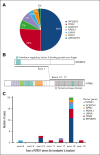
Non-Langerhans cell histiocytoses are a diverse group of histiocytic diseases. Different entities are defined based on clinical, histopathologic, and/or molecular characteristics. This study aimed to define NTRK-rearranged histiocytosis. Through international collaboration, we investigated 50 cases of histiocytosis with pan-tropomyosin receptor kinase (pan-TRK) expression and/or in-frame NTRK rearrangement. We also analyzed 45 control xanthogranulomas using pan-TRK immunohistochemistry and targeted RNA sequencing. Slides were centrally reviewed; clinical and molecular data were collected. The 50 cases comprised 30 children and 20 adults with a median age of 11.5 years (range, 0-73 years) and a male predominance (64%). Most patients (88%) had disease limited to the skin, including a single skin nodule in 41 patients and multiple skin lesions in 3 others. Four newborns presented with skin lesions, hepatomegaly, and thrombocytopenia that required transfusions. The 2 remaining patients had life-threatening lesions of the brain or bronchus. All cases displayed xanthogranuloma histology, often including foamy histiocytes and Touton giant cells. Histiocytes stained positive for pan-TRK in 50 of 50 cases, whereas all 45 control xanthogranulomas without in-frame NTRK fusions stained negative. NTRK1 fusion partners included IRF2BP2 (23/46), TPM3 (12/46), SQSTM1 (3/46), PRDX1 (3/46), NPM1 (2/46), LMNA (2/46), and ARHGEF2 (1/46). Clinical outcomes were favorable, including spontaneous disease regression in 3 of 4 newborns with systemic disease, and rapid clinical response in both patients with a brain or bronchial tumor treated with the TRK inhibitor larotrectinib. This study advances the molecular characterization of histiocytoses and may guide the diagnosis and personalized treatment of patients.
© 2025 American Society of Hematology. Published by Elsevier Inc. Licensed under Creative Commons Attribution-NonCommercial-NoDerivatives 4.0 International (CC BY-NC-ND 4.0), permitting only noncommercial, nonderivative use with attribution. All other rights reserved.
Conflict of interest statement
Conflict-of-interest disclosure: The authors declare no competing financial interests.
Figures


References
-
- Kemps PG, Kester L, Scheijde-Vermeulen MA, et al. Demographics and additional haematologic cancers of patients with histiocytic/dendritic cell neoplasms. Histopathology. 2024;84(5):837–846. - PubMed
-
- So N, Liu R, Hogeling M. Juvenile xanthogranulomas: examining single, multiple, and extracutaneous presentations. Pediatr Dermatol. 2020;37(4):637–644. - PubMed
-
- Song M, Kim SH, Jung DS, Ko HC, Kwon KS, Kim MB. Structural correlations between dermoscopic and histopathological features of juvenile xanthogranuloma. J Eur Acad Dermatol Venereol. 2011;25(3):259–263. - PubMed
-
- Peruilh-Bagolini L, Silva-Astorga M, Hernández San Martín MJ, et al. Dermoscopy of juvenile xanthogranuloma. Dermatology. 2021;237(6):946–951. - PubMed
MeSH terms
Substances
Grants and funding
LinkOut - more resources
Full Text Sources
Miscellaneous

