. 2025 May 13;19(18):17606-17626.
doi: 10.1021/acsnano.5c01184. Epub 2025 May 2.

Nicotinamide Adenine Dinucleotide-Loaded Lubricated Hydrogel Microspheres with a Three-Pronged Approach Alleviate Age-Related Osteoarthritis

Affiliations

Nicotinamide Adenine Dinucleotide-Loaded Lubricated Hydrogel Microspheres with a Three-Pronged Approach Alleviate Age-Related Osteoarthritis

Yanpeng Lin et al. ACS Nano. .

Abstract

Chondrocyte senescence, synovitis, and decreased level of lubrication play pivotal roles in the pathogenesis of age-related osteoarthritis (AROA). However, there are currently no effective therapeutic interventions capable of altering the progression of OA until it reaches advanced stages, necessitating joint replacement. In this study, lubricious and drug-loaded hydrogel microspheres were designed and fabricated by utilizing microfluidic technology for radical polymerization of chondroitin sulfate methacrylate and incorporating nicotinamide adenine dinucleotide (NAD)-loaded liposomes modified with lactoferrin that are positively charged. Mechanical, tribological, and drug release analyses demonstrated enhanced lubrication properties and an extended drug dissemination time for the NAD@NPs@HM microspheres. In vitro assays unveiled the ability of NAD@NPs@HM to counteract chondrocyte senescence. RNA sequencing analysis, untargeted metabolomics analysis, and in vitro experiments on macrophages revealed that NAD@NPs@HM can regulate the metabolic reprogramming of synovial macrophages, promoting their repolarization from the M1 to M2 phenotype, thereby alleviating synovitis. Intra-articular injection of NAD@NPs@HM in aged mice reduced the mechanisms associated with AROA. These results suggest that NAD@NPs@HM may provide extended drug release, improved joint lubrication leading to better gait, and attenuation of AROA pathogenic processes, indicating its potential as a therapeutic approach for AROA.

Keywords: age-related osteoarthritis; cellular senescence; lubrication; macrophage reprogramming; nicotinamide adenine dinucleotide.


Conflict of interest statement

The authors declare no competing financial interest.

Figures
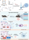
Figure 1
Figure 1
Synthesis protocols and underlying mechanisms of NAD@NPs@HM. (A) The composition of NAD@NPs. (B) Synthesis process of ChSMA. (C) Preparation of NAD@NPs@HM by microfluidic device and photopolymerization process. (D) Mechanistic schema delineating the therapeutic modulation of boundary lubrication, chondrocyte senescence, and macrophage polarization for OA intervention (Created in BioRender. Wu, H. (2025) https://BioRender.com/rr8tovk).
Figure 2
Figure 2
Preparation and characterization of NAD@NPs@HM. (A) A TEM image and the particle size for the blank NPs. (Scale bar: 100 nm). (B) A TEM image and the particle size for the NAD@NPs. (Scale bar: 100 nm). (C) The zeta potential for the blank NPs and the NAD@NPs. (D) 1H NMR spectrum of hydrogel microsphere composition. (E, G) Bright-field plot of NPs@HM, a fluorescent image of Dil-labeled NPs@HM, and a TEM image of NPs@HM. (Scale bar: 25 μm). (H) The particle size for the hydrogel microspheres. (I, J) COF analysis of PBS, blank HM, and NPs@HM. (K, L) Quantification of intracellular NAD(H) level, NAD+/NADH ratio, ATP level, and cellular uptake fluorescent images in chondrocytes and macrophages (scale bar: 50 μm). (M) Drug release profile of NAD@NPs@HM. (N) Degradation curve of NAD@NPs@HM. Data from ≥3 independent experiments (mean ± SD) were analyzed using one-way ANOVA: *P < 0.05, **P < 0.01, ***P < 0.001, ****P < 0.0001.
Figure 3
Figure 3
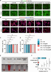
In vitro evaluation of NAD@NPs@HM biocompatibility. (A) Viability assessment of ATDC5 chondrocytes and RAW264.7 macrophages in LPS-stimulated models following 24 h culture with hydrogel microsphere formulations (live/dead staining; scale bar: 500 μm). (B) Cytoskeletal organization in microsphere-treated cells (scale bar: 100 μm). (C, D) Quantitative viability analysis. (E) Hemocompatibility evaluation via erythrocyte incubation assay (2 h coculture). (F) Hemolytic ratio quantification. All experiments were performed in triplicate (n = 3). Data represent mean ± SD (*P < 0.05, **P < 0.01, ***P < 0.001, ****P < 0.0001).
Figure 4
Figure 4
NAD@NPs@HM attenuates chondrocyte senescence and enhances regeneration in vitro. (A, B) Edu staining and wound closure of cells cocultured with different hydrogel microspheres. (Scale bar: 500 μm). (C, D) SA-β gal staining and Toluidine blue staining of ATDC5 cell line. (Scale bar: 200 μm). (E) ROS positive cells in ATDC5 chondrocyte cell line. (Scale bar: 500 μm). (F) The expression of chondrocyte anabolic metabolism, catabolic metabolism, and cellular senescence related genes. (G) Statistical quantification of the EdU positive cells. (H) Quantification of the β-gal positive cells. (I) Quantification of the ROS positive cells in ATDC5 chondrocyte cell line. All experiments were performed in triplicate (n ≥ 3). Data represent mean ± SD (*P < 0.05, **P < 0.01, ***P < 0.001, ****P < 0.0001).
Figure 5
Figure 5
NAD@NPs@HM regulates inflammation in LPS-stimulated macrophages. (A) EdU staining in RAW264.7 cell line after coculture with different hydrogel microspheres in LPS-mediated cellular inflammation model (scale bar: 200 μm). (B) Transwell assay in RAW264.7 cell line. (Scale bar: 500 μm). (C) ROS positive cells in RAW264.7 cell line. (Scale bar: 500 μm). (D) Flow cytometry detection of cell apoptosis. (E) Flow cytometry detection of macrophage types. (F, G) The expression of M1 or M2 macrophage marker genes. All experiments were performed in triplicate (n ≥ 3). Data represent mean ± SD (*P < 0.05, **P < 0.01, ***P < 0.001, ****P < 0.0001).
Figure 6
Figure 6
NAD@NPs@HM regulates the transcriptomics and metabolomics of macrophages stimulated with LPS. (A–C) Volcano plot of differentially expressed genes. (D–F) GO enrichment analysis of transcriptional alterations. (G) Venn diagram related to genes involved in cellular metabolic processes. (H) The expression of the top 15 hub genes in the network. (I) Molecular interactions between NAD and c-Src, including binding interface and allosteric modulation sites. (J) Metabolite set enrichment analysis among the groups. (K) Analysis of the top 15 differential metabolites among the groups.
Figure 7
Figure 7
Gait analysis for the mice after intra-articular injection with hydrogel microspheres. (A) Methodological workflow for establishing the AROA murine model (Created in BioRender. Wu, H. (2025) https://BioRender.com/rr8tovk). (B) Representative gait screen shot and gait intensity. (C) Quantification of Rear Left limb average print area. (D) Quantification of Rear Left limb gait angle. (E) Quantification of Rear Left limb average stride length. (F) Quantification of Rear Left limb average brake time. (G) Quantification of Rear Left limb average stride time. (H) Quantification of Rear Left limb average stance time. Data are represented as means ± SD (n = 6). FR: front right limb. FL: front left limb. RR: rear right limb. RL: rear left limb.
Figure 8
Figure 8
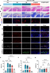
NAD@NPs@HM attenuates AROA progression. (A) Histomorphological micrographs illustrate H&E-stained articular cartilage sections (scale bar: 50 μm). (B) Safranin O-fast green dual staining of subchondral bone demonstrated proteoglycan deposition (red) and osseous matrix (green). Scale bar: low-magnification (100 μm); high-magnification (50 μm). (C) Multiplex immunofluorescence analysis of MMP13 (catabolic marker) and COL2A1 (anabolic marker) with DAPI nuclear counterstaining evaluated cartilage matrix homeostasis. Scale bar: 50 μm. (D) Immunostaining staining for P53, IL-6, and DAPI for assessing the senescence-associated secretory phenotype of cartilage. Scale bar: 50 μm. (E) Microcomputed tomography reconstructions illustrating medial subchondral bone microarchitecture. Scale bar: 1 mm. (F) OARSI-modified Mankin scores of articular cartilage. (G) The hyaline-calcified cartilage (HC/CC) zonal proportion within articular cartilage matrix. (H) Comparative morphometric quantification of trabecular pattern factor (Tb.pf) across subchondral bone microarchitecture was conducted in the following experimental cohorts: 3 month controls (3M), age-matched 18 month groups (18M), and therapeutic intervention arms (18 M + HM, 18 M + NPs@HM, 18 M + NAD@NPs@HM). Number of repetitions of all experiments n ≥ 3. Data are presented as the mean ± SD (*P < 0.05; **P < 0.01; ***P < 0.001 and ****P < 0.0001).
Figure 9
Figure 9
NAD@NPs@HM regulates the inflammatory microenvironment of knee joints in AROA. (A, B) Representative images of HE and Masson staining staining the of synovium. Scale bar: 50 μm. (C) Immunostaining for iNOS and DAPI for assessing the synovitis. Scale bar: 50 μm. (D) Immunostaining for the M1 and M2 types of synovial macrophages. Scale bar: 50 μm. (E) Quantitative analysis of iNOS expression levels in synovial membranes. (F, G) Statistical analysis for the number of CD86+ macrophages and CD206+ macrophages within the synovial membranes of the 3 M group, 18 M group, 18 M + HM group, 18 M + NPs@HM group, and 18 M + NAD@NPs@HM group. Quantitative data were expressed as mean ± SD (n = 3). (*P < 0.05; **P < 0.01; ***P < 0.001 and ****P < 0.0001).

Similar articles

Cited by

References

    1. Johnson V. L.; Hunter D. J. The epidemiology of osteoarthritis. Best Pract. Res. Clin. Rheumatol. 2014, 28 (1), 5–15. 10.1016/j.berh.2014.01.004. - DOI - PubMed
    1. Loeser R. F.; Goldring S. R.; Scanzello C. R.; Goldring M. B. Osteoarthritis: a disease of the joint as an organ. Arthritis Rheum 2012, 64 (6), 1697–1707. 10.1002/art.34453. - DOI - PMC - PubMed
    1. Carr A. J.; Robertsson O.; Graves S.; Price A. J.; Arden N. K.; Judge A.; Beard D. J. Knee replacement. Lancet 2012, 379 (9823), 1331–1340. 10.1016/S0140-6736(11)60752-6. - DOI - PubMed
    1. Bijlsma J. W.; Berenbaum F.; Lafeber F. P. Osteoarthritis: an update with relevance for clinical practice. Lancet 2011, 377 (9783), 2115–2126. 10.1016/S0140-6736(11)60243-2. - DOI - PubMed
    1. He S.; Sharpless N. E. Senescence in health and disease. Cell 2017, 169 (6), 1000–1011. 10.1016/j.cell.2017.05.015. - DOI - PMC - PubMed

Publication types