. 2025 May 19;64(19):9684-9702.
doi: 10.1021/acs.inorgchem.5c00896. Epub 2025 May 2.

Protoporphyrin IX-Derived Ruthenium(II) Complexes for Photodynamic Therapy in Gastric Cancer Cells

Affiliations

Protoporphyrin IX-Derived Ruthenium(II) Complexes for Photodynamic Therapy in Gastric Cancer Cells

Andrés Restrepo-Acevedo et al. Inorg Chem. .

Abstract

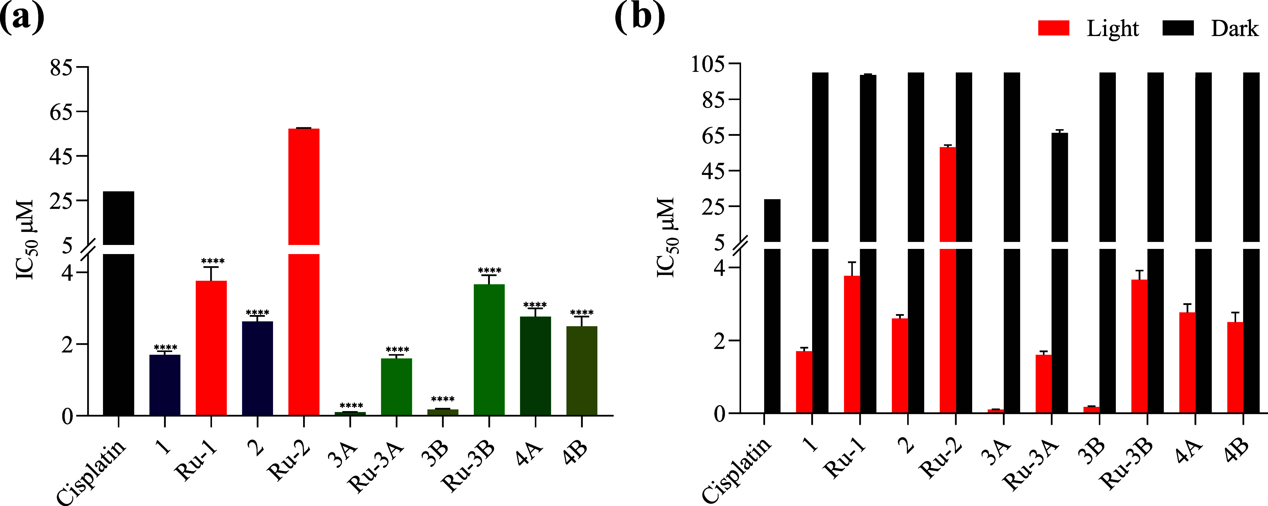
In recent years, photodynamic therapy (PDT) has emerged as a promising alternative to classical chemotherapy for treating cancer. PDT is based on a nontoxic prodrug called photosensitizer (PS) activated by light at the desired location. Upon irradiation, the PS reacts with the oxygen present in the tumor, producing cytotoxic reactive oxygen species (ROS). Compounds with highly conjugated π-bond systems, such as porphyrins and chlorins, have proven to be excellent light scavengers, and introducing a metal atom in their structure improved the generation of ROS. In this work, a series of tetrapyrrole-ruthenium(II) complexes derived from protoporphyrin IX and the commercial drug verteporfin were designed as photosensitizers for PDT. The complexes were almost nontoxic on human gastric cancer cells under dark conditions, revealing remarkable cytotoxicity upon irradiation with light. The ruthenium atom in the central cavity of the chlorin ligand allowed combined mechanisms in photodynamic therapy, as both singlet oxygen and superoxide radicals were detected. Additionally, one complex produced large amounts of singlet oxygen under hypoxic conditions. Biological assays demonstrated that the ruthenium derivatives caused cell death through a caspase 3 mediated apoptotic pathway and via CHOP, an endoplasmic reticulum stress-inducible transcription factor involved in apoptosis and growth arrest.


Conflict of interest statement

The authors declare no competing financial interest.

Figures

Figure 1
Figure 1
Compounds approved or in clinical trials as photosensitizers for PDT.
Scheme 1
Scheme 1. Synthesis of Ligands and Numbering Used for NMR Assignation
Scheme 2
Scheme 2. Synthesis of Ruthenium Complexes and Numbering Used for NMR Assignation
Figure 2
Figure 2
ORTEP diagrams (a) Ru-1 and (b) 3B. Thermal ellipsoids are drawn with a 40% probability level. Hydrogen atoms are omitted for clarity, except for N–H hydrogen atoms.
Figure 3
Figure 3
Absorption spectra of (a) porphyrins and metaloporphyrins and (b) chlorins and metalochlorins in DMSO, 1 × 10⁻⁵ M at 37 °C.
Figure 4
Figure 4
Stability assay for Ru-1 in (a) DMSO and (b) PBS/DMSO 0.1%. Inset: plot of absorbance vs time.
Figure 5
Figure 5
Photodegradation of compound Ru-1 (a, c) and Ru-3B (b, d) in DMSO and PBS/DMSO (0.1%) upon irradiation with white light (50% intensity of a 12 W lamp). Inset: plot of absorbance vs time.
Figure 6
Figure 6
Absorption changes during the determination of singlet oxygen quantum yield of Ru-1 (a) and 3B (b) using DPBF as singlet oxygen trapping in DMSO equilibrated with air (inset: plot of absorbance at 214 nm vs irradiation time). (c) Reaction between DPBF and photosensitizer in the presence of light and oxygen. (d) Percentage singlet oxygen formation quantum yield relative to verteporfin (ΦΔ = 0.77). [PS] = 7 × 10⁻⁶ M; [DPBF] = 2 × 10⁻⁵ M, N = 3.
Figure 7
Figure 7
EPR spectra upon irradiation with white light of the PS in the presence of TEMP spin trap in ethanol/DMSO at room temperature: (a) 1, (b) Ru-1, (c) 3B, and (d) Ru-3B. (e) TEMPO signal intensity after 15 min of irradiation. [PS] = 1 × 10⁻³ M, [TEMP] = 4 × 10⁻³ M, blanc = spectra of TEMP without PS.
Figure 8
Figure 8
EPR spectra upon irradiation for 5 and 15 min of Ru-1 in the presence of TEMP varying the oxygen concentration: (a) 5% of oxygen, (b) 45% of oxygen, (c) 96% of oxygen. (d) TEMPO signal intensity for compounds 1, Ru-1, 3B, and Ru-3B at 15 min of irradiation at different oxygen concentrations. [PS] = 1 × 10⁻³ M, [TEMP] = 4 × 10⁻³ M, blanc = spectra of TEMP without PS.
Figure 9
Figure 9
EPR spectra in methanol/DMSO at room temperature: (a) DMPO in the dark (solid line) and upon irradiation (dotted line), (b) Ru-3A in the dark (black) and upon irradiation (green), and (c) Ru-3B dark (black) and upon irradiation (green). (d) DMPO- O2•–signal intensity after 15 min of irradiation. [PS] = 1 × 10⁻³ M, [DMPO] = 0.07 M. Irradiation time = 15 min.
Figure 10
Figure 10
(a) Graphs showing the mean IC50 inhibitory concentrations presented in Table 2. Student’s t-test is considered significant at p < 0.5. * < 0.05, ** < 0.01, *** < 0.001, **** < 0.0001 with respect to cisplatin. (b) Comparison of the minimum inhibitory concentration in the dark and activated under irradiation with white light for 15 min.
Figure 11
Figure 11
Effects of photoactivation of 1, Ru-1, 3B, and Ru-3B on protein expression on different cell death markers in AGS gastric cancer cells. AGS cells were treated with 1, Ru-1, 3B, Ru-3B, PpIX, or VP at IC50 concentrations for 4 h and then either irradiated with white light or kept in the dark for 15 min and then further cultivated for 24 h in the dark. Protein extraction was performed, and expression of the different markers was determined by Western blot. GAPDH was used as a loading control. Representative Western blot of three independent experiments. ctl = control, untreated cells.
Figure 12
Figure 12
Effects of photoactivation of 1, Ru-1, 3B and Ru-3B on protein expression of ferroptosis and inflammation markers in AGS gastric cancer cells. AGS cells were treated with 1, Ru-1, 3B, Ru-3B, PpIX, or VP at IC50 concentrations for 4 h and then either irradiated with white light or kept in the dark for 15 min and then further cultivated for 24 h in the dark. (a) Expression of the different markers was determined by Western blot. GAPDH was used as a loading control. Representative Western blot of three independent experiments. ctl = control, untreated cells. Graphs represent the quantification of three independent experiments of GPX4 (b) and COX2 (c) expression, compared to not-treated AGS cells (ctl) and normalized to GAPDH expression. Bars above the colons indicate the comparison of dark versus light for each compound. The dark and light results of each compound were compared to those of the respective untreated control condition (ctl). Statistical analysis: 2-way Anova *<0.05, **<0.01, ***<0.001, ns = not-significant.

Similar articles

References

    1. Oluwajembola A. M.; Cleanclay W. D.; Onyia A. F.; Chikere B. N.; Zakari S.; Ndifreke E.; De Campos O. C. Photosensitizers in photodynamic therapy: An advancement in cancer treatment. Results Chem. 2024, 10, 10171510.1016/j.rechem.2024.101715. - DOI
    1. Zhang Y.; Doan B. T.; Gasser G. Metal-Based Photosensitizers as Inducers of Regulated Cell Death Mechanisms. Chem. Rev. 2023, 123 (16), 10135–10155. 10.1021/acs.chemrev.3c00161. - DOI - PubMed
    1. Alderden R. A.; Hall M. D.; Hambley T. W. The Discovery and Development of Cisplatin. J. Chem. Educ. 2006, 83 (5), 72810.1021/ed083p728. - DOI
    1. Johnstone T. C.; Suntharalingam K.; Lippard S. J. The Next Generation of Platinum Drugs: Targeted Pt(II) Agents, Nanoparticle Delivery, and Pt(IV) Prodrugs. Chem. Rev. 2016, 116 (5), 3436–3486. 10.1021/acs.chemrev.5b00597. - DOI - PMC - PubMed
    1. Kelland L. The resurgence of platinum-based cancer chemotherapy. Nat. Rev. Cancer 2007, 7 (8), 573–584. 10.1038/nrc2167. - DOI - PubMed

MeSH terms