. 2025 May 20;6(5):102095.
doi: 10.1016/j.xcrm.2025.102095. Epub 2025 May 1.

Dissecting the immune landscape in pediatric high-grade glioma reveals cell state changes under therapeutic pressure

Affiliations

Dissecting the immune landscape in pediatric high-grade glioma reveals cell state changes under therapeutic pressure

Jenna J LaBelle et al. Cell Rep Med. .

Abstract

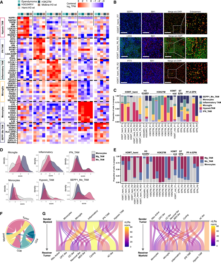
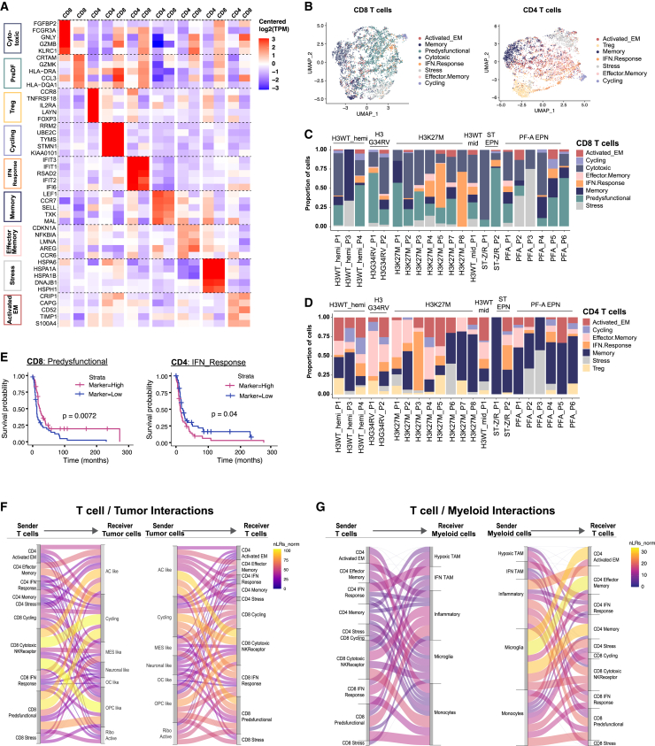
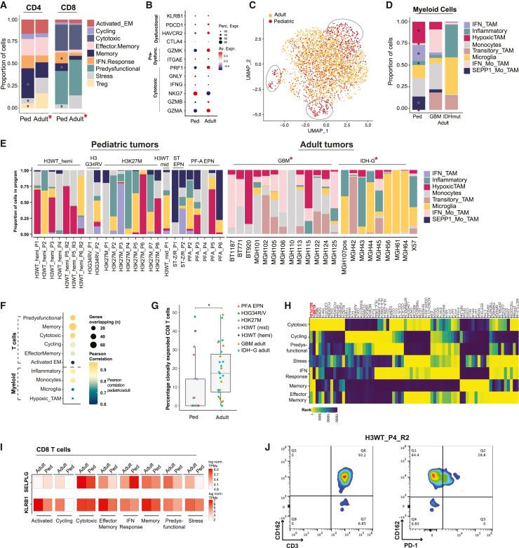
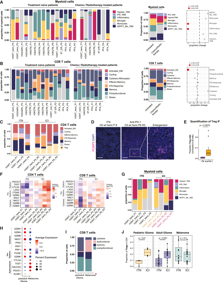
Pediatric high-grade gliomas (pHGGs) are among the most lethal childhood tumors. While therapeutic approaches were largely adapted from adult treatment regime, significant biological differences between pediatric and adult gliomas exist, which influence the immune microenvironment and may contribute to the limited response to current pHGG treatment strategies. We provide a comprehensive transcriptomic analysis of the pHGG immune landscape using single-cell RNA sequencing and spatial transcriptomics. We analyze matched malignant, myeloid, and T cells from patients with pediatric diffuse high-grade glioma (HGG) or high-grade ependymoma, examining immune microenvironment distinctions after chemo-/radiotherapy, immune checkpoint inhibition treatment, and by age. Our analysis reveals differences in the proportions of pediatric myeloid subpopulations compared to adult counterparts. Additionally, we observe significant shifts toward immune-suppressive environments following cancer therapy. Our findings offer valuable insights into potential immunotherapy targets and serve as a robust resource for understanding immune microenvironmental variations across HGG age groups and treatment regimens.

Keywords: immuno-oncology; pediatric high-grade glioma; single-cell RNA-seq; tumor microenvironment; tumor-infiltrating immune cell biology.


Conflict of interest statement

Declaration of interests M.G.F. is a consultant for Twentyeight-Seven Therapeutics and Blueprint Medicines. K.W.W. is a co-founder member of Immunitas Therapeutics. K.W.W. serves on the scientific advisory board of TCR2 Therapeutics, T-Scan Therapeutics, SQZ Biotech, DEM BioPharma, Bisou Bioscience Company, and Nextech Invest and received sponsored research funding from Novartis.

Figures

None
Graphical abstract
Figure 1
Figure 1
Cohort of pHGG-associated immune and malignant cells (A) Clinical and molecular characteristics of the pHGG cohort. Patient samples (n = 31) were profiled after enrichment of CD45-positive cells, followed by Smart-Seq2 (left) or 10× Genomics scRNA-seq (right) from fresh isolated tissue. For patients treated with anti-PD-1, R2 denotes first tumor recurrence, and R3 denotes the following recurrence. Asterisk denotes scRNA-seq tumor data previously published from our laboratory (patients with DMG and ependymoma from the study by Liu et al. and Gojo et al.,, respectively). (B) Representative images of detected malignant/non-immune, myeloid, and T cell assignments in primary tumor sections after spatial transcriptome analysis. Proportions of non-immune and immune cells within pHGGs are indicated. Scale bar is 1 mm for displayed tumor section and 200 μm for selected regions. (C) Schematic depicting the workflow of immune cell and malignant cell enrichment via flow cytometry and downstream analysis.
Figure 2
Figure 2
Myeloid cell expression programs and ligand-receptor interactome (A) Heatmap depicting the relative expression of top marker genes for each myeloid program in the respective pHGG subtype. Transcripts per million (TPM) values were pseudobulked by program (left) and subtype (top), log transformed, and centered by gene. (B) Multiplex IHC staining of myeloid cell markers for identification of subpopulations (general myeloid cell marker: IBA1), hypoxic TAM (marker: LDHA), SEPP1-Mo TAM (marker: SEPP1), and IFN-TAM (marker: IFIT3) on the protein level in pHGG samples. Scale bar, 300 μm. (C) Proportion of each myeloid subpopulation by pHGG subtype. (D) Density plots showing scores for Mg-TAM, Mo-TAM, and monocyte signatures, as described, for each of the six pediatric myeloid subpopulations. (E) Proportions of cells with indicated expression signatures from (D) per pHGG sample. (F) Number of ligand-receptor interactions between tumor cells, myeloid cells, and T cells predicted by CellChat. Thickness of lines denotes unique interaction proportions among all identified potential interactions (determined sample- and program-wise). (G) Ligand-receptor interactions of indicated cell types and subpopulations.
Figure 3
Figure 3
T cell subpopulations in pHGG and their ligand-receptor interactome (A) Heatmap depicting the relative expression of the top marker genes for each T cell program, after merging the pediatric/adult shared programs. TPM values were pseudobulked by program (left) and CD4/CD8 annotation (top), log transformed, and centered by gene. (B) Uniform manifold approximation and projection plots of CD4+ and CD8+ T cells identified via SS2 in this pHGG cohort, colored by their identified T cell program. (C and D) Proportion of each T cell subpopulation, represented by the respective expression program, in our pHGG cohort. (E) Kaplan-Meier survival analysis based on bulk RNA-seq data obtained from the CBTN. The sequencing data were deconvoluted to predict the proportion of CD4+ T cells, CD8+ T cells, myeloid cell, and tumor cell states from our analysis, using Cibersort. Samples were split into “high” proportion and “low” (top/bottom quantiles) and run to determine effects on survival. See Table S5 for survival analysis results for all programs. (F and G) Ligand-receptor interactions of indicated cell types and subpopulations, as in Figure 2G. Thickness of the connecting lines represents the proportion among all identified potential interactions.
Figure 4
Figure 4
Integrative comparison of myeloid and T cells across pediatric and adult HGG (A) Proportion of each T cell subpopulation by age group and CD4/CD8 annotation. Asterisks denote significant changes in observed proportion (p ≤ 0.05). Adult tumor-associated myeloid cells from the study by Neftel et al. and Venteicher et al., (B) Average marker gene expression from dysfunctional, predysfunctional program-associated, and cytotoxic CD8+ T cells in indicated age groups. (C) UMAP of all myeloid cells, colored by age group (adult tumor-associated myeloid cells from the study by Neftel et al. and Venteicher et al.23,24). No integration by Harmony was performed here. (D) Proportion of each myeloid subpopulation by age group and subtype. Asterisks denote significant observed changes in proportion compared to pediatric patients (p ≤ 0.05). Statistical significance assessed using the propeller function from the R package speckle. (E) Proportion of myeloid cell subpopulations, represented by each individual expression program, by subtype and age group. Asterisks denote published datasets. (F) Pearson correlation and the number of overlapping genes between programs shared by adult and pediatric T cells (top) and myeloid cells (bottom). (G) Percentage of clonally expended T cells detected per sample in our pHGG and adult HGG cohort. Asterisk denotes significant change assessed via Student’s t test. Data are represented as mean ± SEM. (H) Heatmap depicting T cell-specific genes ranked according to their expression value among all detected transcripts detected in T cells in the pHGG cohort. (I) Relative expression of SELPLG and KLRB1 mRNAs in T cells from our pHGG and adult HGG cohorts. (J) Flow cytometric analysis for CD162 and PD-1 expression on the surface of T cells (CD3+) from a representative sample from this pHGG cohort.
Figure 5
Figure 5
Chemo-/radiotherapy and ICI immunotherapy affect pHGG immune microenvironment (A) Proportions (left and middle) of myeloid cell subpopulation, represented by each individual program, and quantification of observed changes (right) by treatment status in this pHGG cohort. (B) Proportions (left and middle) of CD8+ T cell subpopulation, represented by each individual program, and quantification of changes (right) by treatment status in this pHGG cohort. Fewer samples due to absent CD8+ T cells in the respective samples. (C) Proportions of CD4+ T cell subpopulations, represented by each individual program, by ICI status in this pHGG cohort. Asterisk denotes significant observed change in proportion (p ≤ 0.05). Statistical significance assessed using the propeller function from the R package speckle. Fewer ITN samples due to absent CD4+ T cells in the respective samples. (D) Multiplex IHC staining for FOXP3 (Treg marker) in samples from ICI-treated and ITN patient (n = 3 each, n images = 12). Scale bar, 300 μm. (E) Quantification of (D): change in observed Treg proportion, normalized to DAPI-positive cells. Student’s t test used to calculate significance. Data are represented as mean ± SEM. (F) Heatmap depicting centered expression values (TPM, transcripts per million) of indicated mRNAs in CD4+ and CD8+ T cells by immunotherapy status. (G) Proportions of myeloid cell subpopulations, represented by each individual program, by immunotherapy status in this pHGG cohort. Colors represent samples from different recurrences in the same patient. (H) Dotplot depicting relative marker gene expression of indicated T cell programs for CD8+ T cells from glioma (our pHGG cohort, adult glioma, and melanoma25). Size of points denote the percentage of cells that the respective gene is expressed in. (I) Proportion of CD8+ T cell subpopulations in same datasets as (H). (J) Proportion of detected CD4+ Treg cells in the indicated cohorts, before and after ICI treatment. Data are represented as mean ± SEM. Significance assessed via Student’s t test.

References

    1. Gröbner S.N., Worst B.C., Weischenfeldt J., Buchhalter I., Kleinheinz K., Rudneva V.A., Johann P.D., Balasubramanian G.P., Segura-Wang M., Brabetz S., et al. The landscape of genomic alterations across childhood cancers. Nature. 2018;555:321–327. doi: 10.1038/nature25480. - DOI - PubMed
    1. Liu I., Jiang L., Samuelsson E.R., Marco Salas S., Beck A., Hack O.A., Jeong D., Shaw M.L., Englinger B., LaBelle J., et al. The landscape of tumor cell states and spatial organization in H3-K27M mutant diffuse midline glioma across age and location. Nat. Genet. 2022;54:1881–1894. doi: 10.1038/s41588-022-01236-3. - DOI - PMC - PubMed
    1. Aggarwal P., Luo W., Pehlivan K.C., Hoang H., Rajappa P., Cripe T.P., Cassady K.A., Lee D.A., Cairo M.S. Pediatric versus adult high grade glioma: Immunotherapeutic and genomic considerations. Front. Immunol. 2022;13 doi: 10.3389/fimmu.2022.1038096. - DOI - PMC - PubMed
    1. Binnewies M., Roberts E.W., Kersten K., Chan V., Fearon D.F., Merad M., Coussens L.M., Gabrilovich D.I., Ostrand-Rosenberg S., Hedrick C.C., et al. Understanding the tumor immune microenvironment (TIME) for effective therapy. Nat. Med. 2018;24:541–550. doi: 10.1038/s41591-018-0014-x. - DOI - PMC - PubMed
    1. Mathewson N.D., Ashenberg O., Tirosh I., Gritsch S., Perez E.M., Marx S., Jerby-Arnon L., Chanoch-Myers R., Hara T., Richman A.R., et al. Inhibitory CD161 receptor identified in glioma-infiltrating T cells by single-cell analysis. Cell. 2021;184:1281–1298.e26. doi: 10.1016/j.cell.2021.01.022. - DOI - PMC - PubMed

MeSH terms