. 2025 May 2;15(1):15420.
doi: 10.1038/s41598-025-98730-5.

Ministring DNA (msDNA): a novel linear covalently-closed DNA with enhanced stability for gene and cell therapy applications

Affiliations

Ministring DNA (msDNA): a novel linear covalently-closed DNA with enhanced stability for gene and cell therapy applications

Wai Kuen Hung et al. Sci Rep. .

Abstract

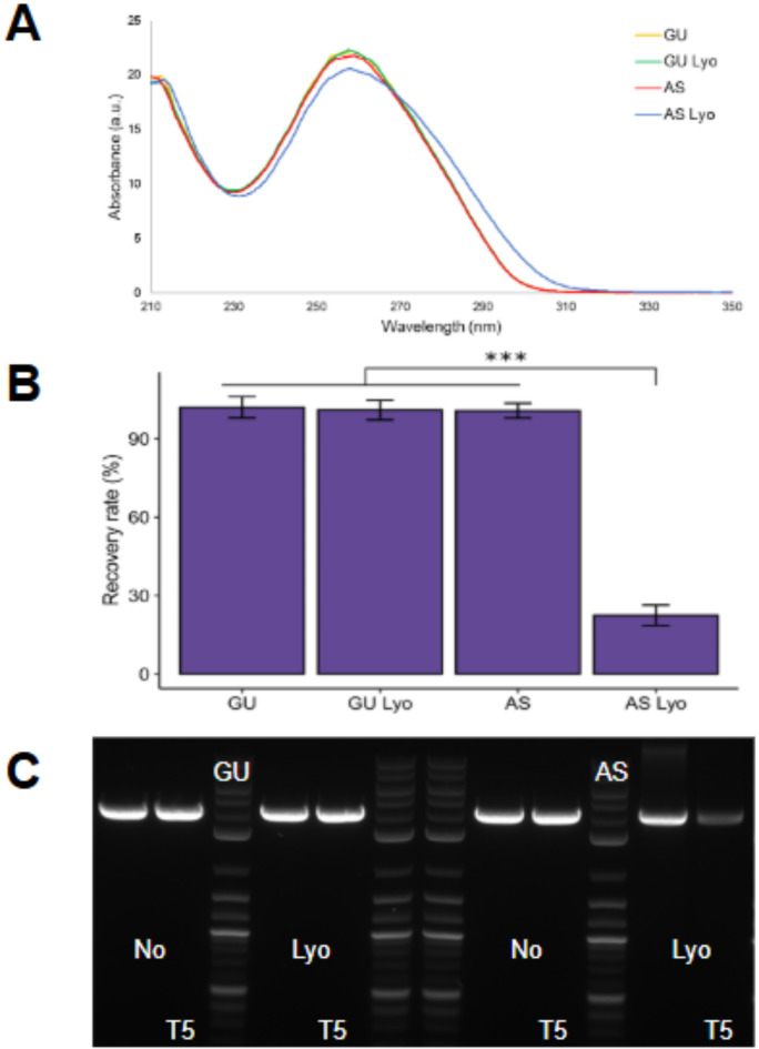
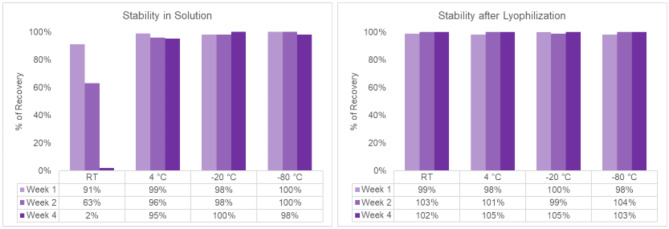
The quality and fidelity of DNA vectors used in genetic medicine and gene therapy either as starting material for manufacturing or as therapeutic ingredients play a critical role in determining ultimate clinical success. Ministring DNA (msDNA), is a novel minivector that is a linear covalently-closed (LCC) double-stranded DNA molecule devoid of immunogenic bacterial sequences (e.g., origin of replication, antibiotic resistant cassette). Similar to traditional plasmids, msDNA is manufactured in vivo in E. coli and therefore benefits from the scalability of E. coli -based systems and the ~ 1000-fold enhanced fidelity conferred by the mismatch repair (MMR) mechanism. In this paper, we address the improved stability of msDNA. We show that due to the torsion-free structure, msDNA is more stable to chemical and mechanical stress than conventional plasmid DNA. Moreover, we demonstrate that lyophilization can further improve the long-term stability of msDNA, reducing the need for cold chain storage. Therefore, we propose that msDNA can be a new paradigm for genetic medicine by offering genetic material with lower immunogenicity, reduced risk of insertional mutagenesis, and higher fidelity and stability.

Keywords: Chemical and mechanical stability; Gene therapy; Keywords; Linear covalently closed DNA; Lyophilization; Ministring DNA.


Conflict of interest statement

Declarations. Competing interests: The authors are employees and own stock in Mediphage Bioceuticals, Inc. Ethical approval: No ethical approval was required as no human or animal experiments we carried out.

Figures

Fig. 1
Fig. 1
The msDNA is a linear covalently-closed double-stranded DNA vector free of bacterial backbone DNA. msDNA carries a minimal expression cassette comprised of the eukaryotic promoter, transgene or genes of interest, and regulatory sequences. The ends of the msDNA are covalently closed by a protelomerase induced hairpin formation. [Image obtained with permission from Mediphage Bioceuticals Inc.]
Fig. 2
Fig. 2
Preliminary stability test of msDNA (Construct M, 3.1 kb, 53% GC) in nuclease-free pharmaceutical grade water. (A) AGE of samples at the start of the experiment (Day 1) and after 3 months of storage, highlighting significant DNA degradation over time at room temperature (23–25 °C). All temperatures are in degrees Celsius (°C). (B) Ultra-high-performance liquid chromatography (UHPLC) traces of the sample at various time intervals (Start, 1 week, 1 month and 3 months). For detailed UHPLC traces at each time point, refer to Figure S1.
Fig. 3
Fig. 3
Stability assessment of msDNA (Construct A, 4.3 kb, 59% GC) and isogenic pDNA under mechanical stress. Both pDNA and msDNA were subjected to mechanical forces by weak (bath; 30–45 watts/liter) and strong (probe-tip; 100,000 watts/liter) sonication. The msDNA demonstrated stability under weak sonication and exhibited greater stability than pDNA under strong sonication for brief exposure times. (A): Agarose gel electrophoresis AGE of pDNA and msDNA following weak and strong sonication and (B): Quantitative analysis of the AGE comparing the stability of msDNA with pDNA (for pDNA only the supercoiled fraction is considered for quantification). Bars show the average of three biological replicates, and error bars show one standard deviation. T-test: ***p < 0.001, *p < 0.05, the statistics data is summarized in Table S2.
Fig. 4
Fig. 4
Stability assessment of msDNA (Construct A, 4.3 kb, 59% GC) and isogenic pDNA under chemical stress following overnight incubation. (A) Quantitative analysis of agarose gel electrophoresis comparing the stability of msDNA with pDNA (note: only the supercoiled fraction of the pDNA is considered for analysis). Bars show the average of three biological replicates, and error bars show one standard deviation. T- test: *p < 0.05,, the statistics data is summarized in Table S3. (B) AGE results of pDNA and msDNA at various pH conditions after approximately 16 h (overnight) of incubation. Both msDNA and pDNA exhibit stability within the pH range of 5 to 10.5 but show signs of degradation at pH 4 or below. Notably, msDNA remains stable at pH 4, while pDNA does not. Note: For pDNA, only the supercoiled fraction (the brightest band between 4–5 kb) is considered for quantification as the other isoforms (e.g., nicked, open circular, and linearized) present in pDNA are fully processed by T5.
Fig. 5
Fig. 5
Stability assessment of msDNA (Construct A, 4.3 kb, 59% GC) to lyophilization at various scales in water. (A): UV-Vis spectra of the various DNA samples before and after lyophilization, with the NO (Control) sample representing the pre-lyophilization sample. The 0.05, 0.1, 0.5 and 1.0 mg indicate the different scales of lyophilization. (B) Quantitative analysis of the recovery following T5-exonuclease treatment observed in the agarose gel electrophoresis (AGE) gel. Bars show the average of three technical replicates, and error bars show one standard deviation. One-way ANOVA with TukeyHSD test: *p < 0.05 (Table S4). (C) AGE gel of the lyophilization samples treated with/without T5 exonuclease.
Fig. 6
Fig. 6
Interactions of msDNA (Construct G, 3.7 kb, 52% GC) with salts prior to lyophilization. (A): UV spectra of samples treated with Guanidine (GU) and Ammonium Sulfate (AS) treated samples before and after lyophilization (after lyophilization indicated by Lyo). The AS sample shows a strong hyperchromic shift post lyophilization. (B) Quantitative analysis of the recovery following T5-exonuclease treatment observed in the agarose gel electrophoresis (AGE) gel. Bars show the average of three technical replicates, and error bars show one standard deviation. One-way ANOVA with TukeyHSD test: ***p < 0.001 (Table S5). (C): AGE image of GU and AS samples treated with T5, assessing integrity after lyophilization.
Fig. 7
Fig. 7
Role of excipient in the stability of msDNA (Construct G, 3.7 kb, 52% GC). (A, B, C): UV-vis spectra of the samples before (blue) and after (orange) lyophilization. (D and E): Quantitative analysis and AGE of the samples before and after lyophilization and their corresponding recovery rates after exposure to T5-exonuclease. The trehalose additive demonstrates the most effective protection against lyophilization-induced damage. Bars show the average of three technical replicates, and error bars show one standard deviation. One-way ANOVA with TukeyHSD test: **p < 0.005 (Table S6).
Fig. 8
Fig. 8
Long term stability of msDNA (Construct A, 4.3 kb, 59% GC) under different storage conditions. At low temperatures (4 °C, -20 °C and − 80 °C), msDNA remains stable in both solution (A) and lyophilized form (B). At room temperature (RT), msDNA is stable in-solution for approximately 1 week only, but it maintains stability in lyophilized form for at least 4 weeks. The raw data supporting this graph can be found in Figures S7-S9. Note: AGE analysis has an error around 5–10% therefore values near 95–105% are considered complete recovery or no loss.

Similar articles

References

    1. Wang, D., Tai, P. W. L. & Gao, G. Adeno-associated virus vector as a platform for gene therapy delivery. Nat. Rev. Drug Discov18, 358–378 (2019). - PMC - PubMed
    1. Yáñez-Muñoz, R. J. et al. Effective gene therapy with nonintegrating lentiviral vectors. Nat. Med.12, 348–353 (2006). - PubMed
    1. Rossor, A. M., Reilly, M. M. & Sleigh, J. N. Antisense oligonucleotides and other genetic therapies made simple. Pract. Neurol.18, 126–131 (2018). - PubMed
    1. Lauffer, M. C., van Roon-Mom, W. & Aartsma-Rus, A. Possibilities and limitations of antisense oligonucleotide therapies for the treatment of Monogenic disorders. Commun. Med.4, 6 (2024). - PMC - PubMed
    1. Weissman, D., Karikó, K. & mRNA Fulfilling the promise of gene therapy. Mol. Ther.23, 1416–1417 (2015). - PMC - PubMed

LinkOut - more resources