

Strong Enhancement of Two-Photon Absorption and Emergence of Unusual Extinction Saturation in Silver Sulfide Quantum Dots Integrated with Gold and Silica Nanostructures
- PMID: 40317259
- PMCID: PMC12086773
- DOI: 10.1021/acsami.5c00984
Strong Enhancement of Two-Photon Absorption and Emergence of Unusual Extinction Saturation in Silver Sulfide Quantum Dots Integrated with Gold and Silica Nanostructures
Abstract
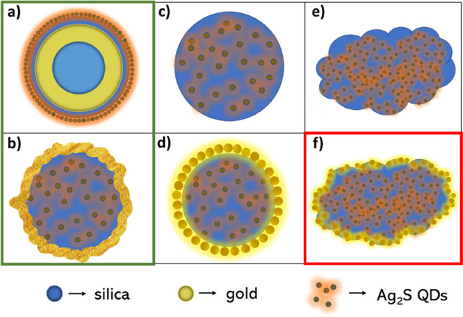
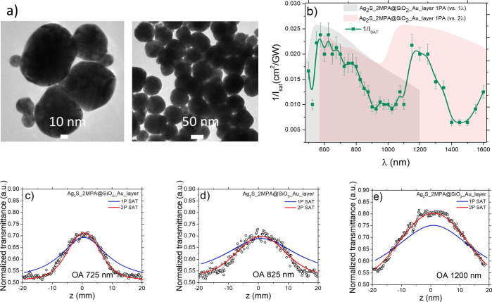
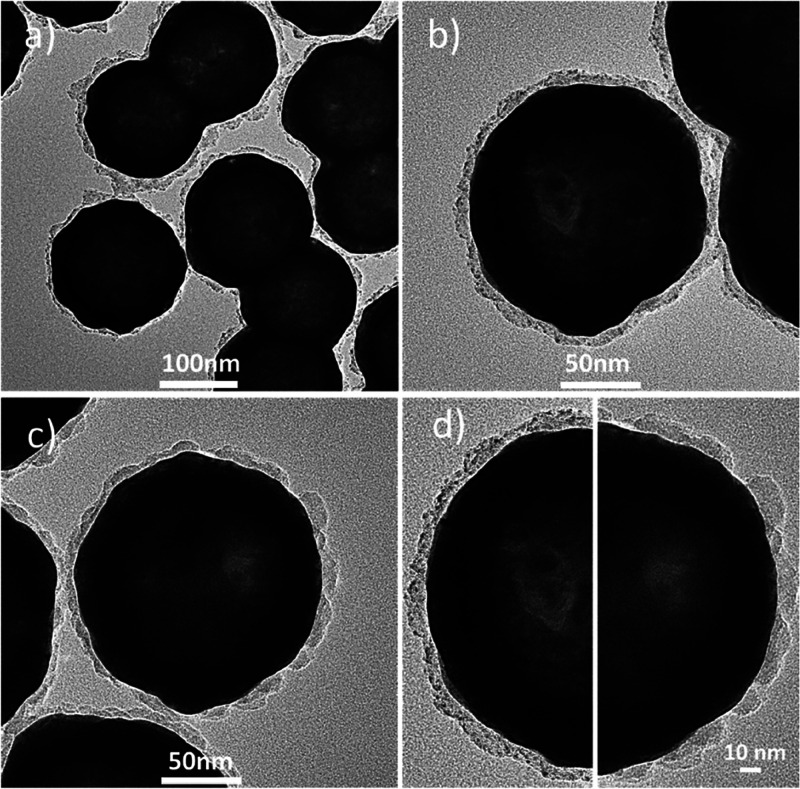
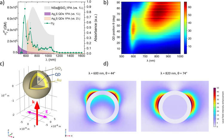
Hybrid nanosystems, such as those combining plasmonic, dielectric, and quantum-confined nanostructures, have long been of interest for enhancing and tailoring diverse light-matter interactions. Here, we present a series of hybrid nanomaterials exhibiting strongly enhanced nonlinear optical (NLO) properties, fabricated by combining silver sulfide quantum dots (Ag2S QDs) with silica and gold nanostructures. We studied their NLO properties (two-photon absorption and saturable absorption) in colloidal solutions over a wide spectral range (500-1600 nm) using the femtosecond Z-scan technique. Embedding Ag2S QDs into silica nanospheres gives rise to remarkable enhancement of two-photon absorption (up to a factor of 16 increase in the merit factor σ2/M compared to bare QDs), whereas covering such QD-doped silica nanospheres with gold nanoparticles or attaching the QDs to the surface of gold nanoshells (NSs) leads to even further enhancement (up to 73-fold increase in σ2/M), accompanied by a competing effect of saturable absorption. Furthermore, in the case of QD-doped silica spheres covered with a continuous gold layer, we observe a previously unreported saturation of extinction in the near-infrared region that follows an unusual intensity dependence, suggesting the involvement of two-photon absorption as the pumping mechanism. In addition to the experimental studies, we have performed numerical simulations, revealing the plasmonic origin of the observed spectral dependences of the NLO properties, with the underlying enhancement mechanisms involving local field enhancement and, possibly, also coupling between plasmon modes and QD excitons, giving rise to a double peak in the σ2 spectrum. Our findings demonstrate the unique potential of hybrid NLO nanomaterials combining quantum-confined, plasmonic, and dielectric components.
Keywords: Ag2S; Z-scan; gold nanoparticles; nanomaterials; nanoshells; plasmon resonance; quantum dots; saturable absorption; silver sulfide; two-photon absorption.
Conflict of interest statement
The authors declare no competing financial interest.
Figures
Similar articles
-
Third-Order Nonlinear Optical Properties of Aqueous Silver Sulfide Quantum Dots.J Phys Chem Lett. 2023 Dec 14;14(49):11117-11124. doi: 10.1021/acs.jpclett.3c02820. Epub 2023 Dec 6. J Phys Chem Lett. 2023. PMID: 38054438 Free PMC article.
-
Strong increase in the effective two-photon absorption cross-section of excitons in quantum dots due to the nonlinear interaction with localized plasmons in gold nanorods.Nanoscale. 2021 Mar 4;13(8):4614-4623. doi: 10.1039/d0nr08893e. Nanoscale. 2021. PMID: 33605966
-
An experimental and theoretical mechanistic study of biexciton quantum yield enhancement in single quantum dots near gold nanoparticles.Nanoscale. 2015 Apr 21;7(15):6851-8. doi: 10.1039/c5nr00274e. Nanoscale. 2015. PMID: 25806486
-
Multifunctional compact hybrid Au nanoshells: a new generation of nanoplasmonic probes for biosensing, imaging, and controlled release.Acc Chem Res. 2014 Jan 21;47(1):138-48. doi: 10.1021/ar400086e. Epub 2013 Aug 30. Acc Chem Res. 2014. PMID: 23992824 Review.
-
Ag2S quantum dot theragnostics.Biomater Sci. 2021 Jan 5;9(1):51-69. doi: 10.1039/d0bm01576h. Biomater Sci. 2021. PMID: 33185212 Review.
References
-
- Gordel-Wójcik M.; Tracz J.; Malik M.; Czeluśniak I.; Zych E. Silver Sulfide Quantum Dots Stabilized with L-Cysteine - Optimization of Synthesis in Aqueous Phase and Optical Characterization. Opt Mater. 2024, 155, 11583110.1016/j.optmat.2024.115831. - DOI
-
- Ibrahim S. K.; Albadr R. J.; Ballal S.; Sur D.; Jie J. C.; Sharma G. C.; Sharma R. S. K.; Bareja L.; Khujanazarov U.; Bhakuni P. N.; athab A. H.; Mansoor A. S.; Radi U. K.; Abd N. S.; Ahmad Z. Advances in Sulfur-Based Quantum Dots for Environmental Sensing: Synthesis, Characterization, Challenges, and Future Prospects. Inorg. Chem. Commun. 2025, 174, 11406410.1016/j.inoche.2025.114064. - DOI
-
- Lims S. C.; Tran N. A.; Dao V.-D.; Pham P. V. The World of Quantum Dot-Shaped Nanoparticles: Nobel Prize in Chemistry 2023: Advancements and Prospectives. Coord. Chem. Rev. 2025, 528, 21642310.1016/j.ccr.2024.216423. - DOI
LinkOut - more resources
Full Text Sources
