Review
. 2025 May 3;44(2):50.
doi: 10.1007/s10555-025-10265-3.

Molecular mechanisms of metastatic peritoneal dissemination in gastric adenocarcinoma

Affiliations
Review

Molecular mechanisms of metastatic peritoneal dissemination in gastric adenocarcinoma

Deanna Ng et al. Cancer Metastasis Rev. .

Abstract

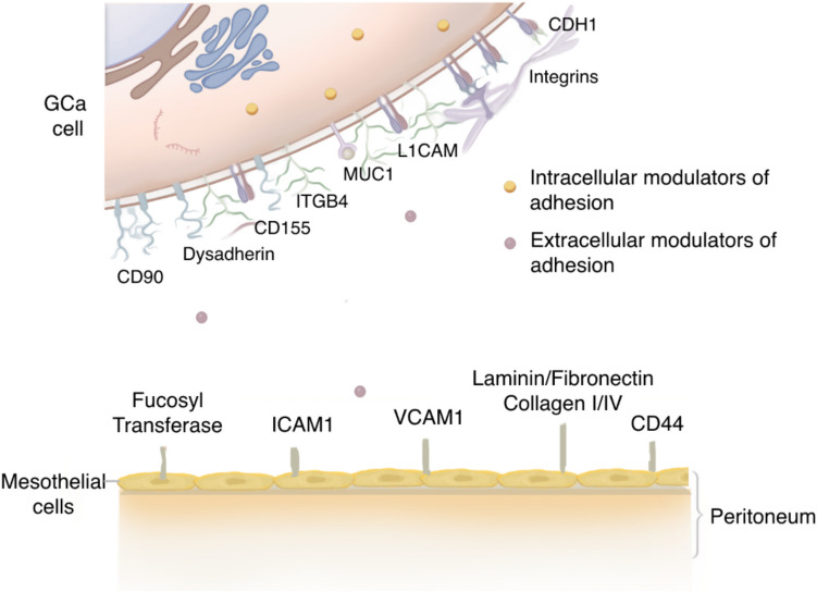
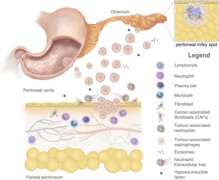
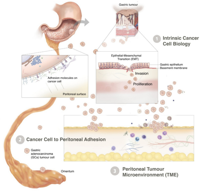
Peritoneal dissemination portends a dismal prognosis in patients with gastric adenocarcinoma in the context of limited effective treatments. The underlying cellular processes that drive gastric peritoneal carcinomatosis remain unclear, limiting the application of novel targeted therapies. In this comprehensive review, we aimed to identify and summarize all existing context-dependent molecular mechanisms that have been implicated in peritoneal dissemination and peritoneal carcinomatosis establishment from primary gastric adenocarcinoma. We applied a multilevel examination including data from in vivo murine models using human gastric cancer cell lines, in vitro technique-based studies, ex vivo models, and genomic/proteomic and molecular profiling analyses to report on various aspects of gastric cancer peritoneal metastasis biology. Mechanisms promoting peritoneal dissemination were grouped into three main functional categories: (1) intrinsic cancer cell biology, (2) cancer cell-peritoneal surface adhesion, and (3) peritoneal tumor microenvironment. We identified significant overlap among the three categories, indicating a complex interplay between multiple molecular mechanisms. By interrupting these pathways, peritoneal-directed therapies have the potential to improve quality and length of life in patients with high-risk primary gastric cancer.

Keywords: Biomarker; Gastric cancer; Microenvironment; Peritoneal carcinomatosis; Tumor progression.


Conflict of interest statement

Declarations. Conflict of interest: The authors declare no competing interests.

Figures

Fig. 1
Fig. 1
Key steps in the gastric cancer peritoneal metastatic cascade. Illustration of the five main processes by which gastric adenocarcinoma cells spread and metastasize within the peritoneal cavity: (1) Invasion through the layers of the gastric wall, into the peritoneal cavity; (2) migration through the peritoneal cavity; (3) adhesion to the mesothelial cell lining of the peritoneal surface; (4) invasion through the basement membrane into the submesothelial layer; (5) growth and angiogenesis
Fig. 2
Fig. 2
PRISMA flowchart of study selection process
Fig. 3
Fig. 3
Mediators and modulators of tumor cell-mesothelial adhesion. Both intra-cellular modulators that alter receptors and proteins on the gastric tumor cell surface and extra-cellular modulators that optimize the structure and environment of the extracellular matrix are involved in gastric cancer cell to peritoneal surface adhesion. GCa = gastric cancer
Fig. 4
Fig. 4
Factors within the tumor microenvironment (TME) that influence gastric cancer peritoneal metastasis. This schematic highlights key components within the TME implicated in gastric cancer peritoneal metastasis. Peritoneal milky spots, lymphoid-rich regions within the greater omentum, serve as major implantation sites for metastases. They consist of mesothelial-covered mesenchymal cells, macrophages, lymphocytes, and mast cells, facilitating tumor cell adhesion. Cancer-associated fibroblasts (CAFs) are a heterogeneous and plastic population of activated fibroblasts within the tumor stroma that support extracellular matrix deposition, remodeling, and reciprocal signaling with cancer cells, thereby enhancing metastasis. Exosomes, vesicles released from gastric cancer cells and omental adipocytes into malignant ascites, promote EMT, invasion, migration, and adhesion. Neutrophil extracellular traps (NETs) consist of condensed extracellular fiber scaffolds that can shield the tumor from host immune responses and promote growth and proliferation. Hypoxia-inducible factor- 1α (HIF- 1α), a transcription factor stabilized under the hypoxic conditions in the peritoneal cavity, regulates cell proliferation, apoptosis, and energy metabolism, contributing to the aggressiveness of gastric cancer cell spread
Fig. 5
Fig. 5
Summary of key processes implicated in peritoneal metastasis from primary gastric cancer. This schematic illustrates the sequential but interconnected steps involved in peritoneal carcinomatosis, with a complex interplay of molecular interactions and tumor microenvironment components. Gastric tumor cells undergo epithelial-mesenchymal transition (EMT), losing epithelial traits and gaining mesenchymal properties that enable invasion and proliferation ((1) intrinsic cancer cell biology). Once in the peritoneal cavity, free-floating cancer cells interact with adhesion molecules on the peritoneal surface, facilitating cancer cell adhesion ((2) cancer cell to peritoneal adhesion). This adhesion is mediated by integrins, selectins, and other molecular regulators that promote tumor anchoring and colonization. The peritoneal tumor microenvironment (TME) consists of immune and stromal components, including cancer-associated fibroblasts (CAFs), tumor-associated macrophages, neutrophils, and lymphocytes, which contribute to tumor progression by remodeling the extracellular matrix, suppressing immune responses, and promoting angiogenesis ((3) peritoneal tumor microenvironment)

Similar articles

References

    1. Pastushenko, I., & Blanpain, C. (2019). EMT transition states during tumor progression and metastasis. Trends in Cell Biology,29(3), 212–226. - PubMed
    1. Hippo, Y., et al. (2001). Differential gene expression profiles of scirrhous gastric cancer cells with high metastatic potential to peritoneum or lymph nodes. Cancer Research,61(3), 889–895. - PubMed
    1. Bai, F. H., et al. (2012). Screening and identification of peritoneal metastasis-related genes of gastric adenocarcinoma using a cDNA microarray. Genetics & Molecular Research,11(2), 1682–1689. - PubMed
    1. Kanda, M., & Kodera, Y. (2016). Molecular mechanisms of peritoneal dissemination in gastric cancer. World Journal of Gastroenterology,22(30), 6829–6840. - PMC - PubMed
    1. Rau, B., et al. (2020). Peritoneal metastasis in gastric cancer: Results from the German database. Gastric Cancer,23(1), 11–22. - PubMed

MeSH terms

LinkOut - more resources