Comparative Study
. 2025 Jul;40(7):1265-1278.
doi: 10.1002/mds.30221. Epub 2025 May 2.

Bilateral Focused Ultrasound Thalamotomy for Essential Tremor: Clinical Outcomes Compared to Bilateral Deep Brain Stimulation and Probabilistic Lesion Mapping

Affiliations
Comparative Study

Bilateral Focused Ultrasound Thalamotomy for Essential Tremor: Clinical Outcomes Compared to Bilateral Deep Brain Stimulation and Probabilistic Lesion Mapping

Can Sarica et al. Mov Disord. 2025 Jul.

Abstract

Background: The efficacy and adverse events (AEs) of bilateral magnetic resonance-guided focused ultrasound (MRgFUS) thalamotomies for essential tremor (ET) have not been compared to those of deep brain stimulation (DBS). Furthermore, it is uncertain whether second-side thalamotomies can be positioned differently from the first without compromising effectiveness.

Objective: We aimed to indirectly compare bilateral MRgFUS and DBS, while identifying optimal lesion/stimulation locations.

Methods: We retrospectively examined 41 ET patients who received either bilateral thalamic DBS (n = 22) or MRgFUS (n = 19) surgery. The primary outcome was the comparison of modalities for change in Clinical Rating Scale for Tremor (CRST) from baseline to post second surgery. We characterized AEs, generated probabilistic maps, and tracked streamlines intersecting lesions. First-side lesions were always intentionally placed ventrally (z = 0/+2 mm above the intercommissural plane [ICP]), and second-side lesions were placed dorsally (z = +3 mm above ICP).

Results: Tremor scores improved significantly after second surgeries (MRgFUS: 56.3 ± 7.1 to 24.2 ± 10.4, P < 0.001; DBS: 58.8 ± 11.6 to 25.0 ± 13, P < 0.05, mean follow-up: 23/26 months), with no differences between modalities. Following first surgeries, scores were MRgFUS: 37.9 ± 7.9 and DBS: 35.2 ± 13.6, with significant improvement from baseline (P < 0.001, mean follow-up: 40/73 months). All AEs were grade 1-2, with AE-free rates of 41% for DBS and 32% for MRgFUS. First-side lesions exhibited maximal efficacy in the ventral Vim, extending to posterior subthalamic area (PSA), whereas second-side lesions demonstrated maximal efficacy in the dorsomedial Vim-Vop border. DBS maps corroborated this finding and confined to Vim-Vop border. Lesions intersecting with networks interconnected with the supplementary motor area, in addition to M1, were associated with improved outcomes.

Conclusions: The efficacies of bilateral MRgFUS and DBS appear comparable. MRgFUS probabilistic maps vary with different targeting methods, revealing two distinct sweet spots: dorsal Vim-Vop border and ventral Vim/PSA. © 2025 The Author(s). Movement Disorders published by Wiley Periodicals LLC on behalf of International Parkinson and Movement Disorder Society.

Keywords: bilateral surgery; deep brain stimulation; fiber filtering; focused ultrasound; probabilistic mapping.


Figures

FIG. 1
FIG. 1
Patient selection and analysis of tremor outcomes and adverse events (AEs) (A) among 189 magnetic resonance‐guided focused ultrasound (MRgFUS) patients, 27 received bilateral treatment; 8 were excluded due to non‐essential tremor (ET) diseases (3 cases: 2 PD, 1 dystonic tremor), incomplete surgery (1 claustrophobia), or missing Clinical Rating Scale for Tremor (CRST) scores (4 cases). Of 194 deep brain stimulation of the ventral intermediate nucleus (Vim‐DBS) patients, 39 had bilateral implantation (36 staged, 3 simultaneous); 17 were excluded for non‐ET diagnoses (10 cases: 3 Parkinson's disease [PD], 2 orthostatic tremor, 2 dystonic tremor, 1 Charcot–Marie‐Tooth, 1 fragileX tremor/ataxia, 1 myoclonus), missing CRST scores (5 cases), and 4 lead implants (2 cases). (B) The treatment demonstrated significant efficacy in both the MRgFUS (F1.77,31.88 = 107.39, P < 0.001, n = 19) and DBS (F1.68,31.98 = 65.01, P < 0.001, n = 20) cohorts. The linear mixed‐effects model revealed significant effects of time (linear: estimate = −23.8649, standard error [SE] = 1.8526, t = −12.882, P < 0.001; quadratic: estimate = 5.4501, SE = 1.8526, t = 2.942, P = 0.00398). However, it showed no significant main effect of intervention or interaction with intervention. (C) The baseline total CRST score for the MRgFUS cohort (56.3 ± 7.6) improved to 37.9 ± 7.1 (P < 0.001) after the first surgery and further improved to 24.2 ± 7.9 (P < 0.001) following the second. (D) First‐side surgeries yielded a 62% improvement in the corresponding lateralized scores in the MRgFUS cohort (t [36]= −13.214, P < 0.001), whereas second‐side surgeries demonstrated a 48% improvement (W = 26.5, P < 0.001). No significant correlation was found between first‐ and second‐side scores (r = 0.16, P = 0.52). (E) In DBS cohort, baseline total CRST score (58.8 ± 11.7) improved to 35.3 ± 11.6 (P < 0.001) after the first surgery, and further to 25.1 ± 13.7 (P = 0.01) after the second surgery. (F) In the DBS cohort, first‐side surgeries resulted in a 58% improvement (t [38]= −8.417, P < 0.001), whereas second‐side surgeries led to a 46% improvement (t [37]= −5.327, P < 0.001). A positive correlation was observed between the changes in first‐side and second‐side lateralized scores (r = 0.51, P < 0.05). (G) Peak AEs were mapped based on their grades and whether they occurred after the first or second surgeries, spanning from first surgery to the latest follow‐up visit. Resolved AEs at any time point prior to the latest follow‐up are indicated with an “X” mark. Among the 22 permanent AEs in the DBS cohort at the last follow‐up, 10 (45%) were not stimulation‐dependent, indicating that turning off the DBS did not alleviate these AEs. These events may be related to the surgery itself or solely due to disease progression, making differentiation challenging. These cases are marked with a “?.” Hardware‐related complications in DBS patients included internal pulse generator discomfort/migration in 3 patients, with surgical intervention for Patient 2 and staged bilateral lead revision in Patient 11 due to reduced treatment effect. Although 5 of 22 (23%) DBS patients experienced falls during the last year of follow‐up, only 1 of 19 (5%) MRgFUS patients did. G1/2, grade 1/2; P‐values: * <0.05, ** < 0.01, *** < 0.001. [Color figure can be viewed at wileyonlinelibrary.com]
FIG. 2
FIG. 2
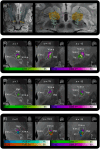
Comparison of first‐side and second‐side magnetic resonance‐guided focused ultrasound (MRgFUS) average n‐p maps and visualization of optimal tremor response areas. (A) Axial (z = 2), coronal (y = −16), and sagittal (x = −14) MRgFUS n‐p maps for the first‐side and second‐side surgeries were superimposed on the 7‐T magnetic resonance imaging (MRI) ex vivo 200‐μm human brain template, along with thalamic nuclei borders derived from the DISTAL atlas version 1.1. All maps and thalamic nuclei were spline interpolated using FSL, with a 10% minimum threshold applied to ensure that the volume matched with that of the non‐interpolated maps. Although the two maps largely overlapped within the ventral intermediate nucleus (Vim), the first‐side map extended into the posterior subthalamic area (PSA), and the second‐side map extended to the dorsal border of the Vim. (B) To visualize optimal tremor response areas, the maps were high‐thresholded at the 80th percentile (arbitrarily chosen) of the maximum mean improvement per voxel score. Scores were rounded to multiples of 5 for easier visualization. Images were presented in axial (z = 3), coronal (y = −16), and sagittal (x = −14) views, respectively. Although the PSA‐extending first‐side lesion map showed optimal tremor response when lesions were centered in Vim externus (V.im.e) and extended into the PSA, the second‐side lesion maps, confined within the thalamus and not extending into the PSA, demonstrated optimal tremor response when lesions were located dorsally at the V.im.e‐Vop border. (C) Additional axial slices of high‐thresholded maps were provided at z = 2.5, 2, 1, 0, −1, and −2.5, all aligned with the same coronal (y = −16) and sagittal (x = −14) planes. [Color figure can be viewed at wileyonlinelibrary.com]
FIG. 3
FIG. 3
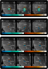
Visualization of deep brain stimulation (DBS) electrode placements and comparison of first‐side and second‐side DBS average n‐p maps, along with all high‐threshold maps. (A) DBS electrode placement in 20 patients were displayed on an axial plane (z = −9.2 mm) of the 7‐T magnetic resonance imaging (MRI) ex vivo 200‐μm human brain template, with a superior view of the Vim, as defined by the DISTAL atlas, version 1.1. Two patients (FUS017 and FUS018) were excluded from the analysis due to the absence of preoperative MRIs. (B) DBS electrode contacts were visualized as point cloud representations. (C) The DBS n‐p maps were displayed on axial (z = 1), coronal (y = −15), and sagittal (x = −15) slices, respectively. The first‐side and second‐side maps showed minimal overlap, both confined to the thalamus and not extending into the posterior subthalamic area (PSA). (D) High‐thresholded DBS maps indicated that the optimal tremor response was located at the Vim externus‐ventro‐oralis posterior (V.im.e‐Vop) border for both sides. However, the second‐side volume of tissue activated (VTAs) were positioned more rostromedially at dorsal levels, whereas the first‐side VTAs were situated more ventrally. (E) All high‐threshold maps (MRgFUS and DBS) were displayed together. Although the maps extended to the dorsal border of the Vim and beyond (with the highest dorsal points at z = 5.5 for second‐side DBS and z = 7.25 for second‐side magnetic resonance‐guided focused ultrasound [MRgFUS]), the high‐threshold maps did not indicate any areas of optimal improvement above z = 4.5. The maps confined to the thalamus—including first‐side and second‐side DBS and second‐side MRgFUS—predominantly overlapped within the V.im.e, between the Vop and Vim externus (V.im.i) and shifted more rostrally at ventral levels. In contrast, the PSA‐extending map (first‐side MRgFUS) exhibited a distinctly different pattern, remaining centrally positioned within the V.im.e more caudally compared to the thalamus‐confined lesions and extending into the PSA. [Color figure can be viewed at wileyonlinelibrary.com]
FIG. 4
FIG. 4
Common and sweet streamlines associated with magnetic resonance‐guided focused ultrasound (MRgFUS) treatment. (A) In the first column, streamlines common to at least 75% of all lesions on one side were selected and displayed on a sagittal slice (x = −24) of the 7‐T ex vivo 200‐μm human brain template. These streamlines were positioned within the MNI coordinates of x = −7 and −23. Common streamlines for the first‐side surgery primarily connected the primary motor cortex (M1) and the posterior supplementary motor area (SMA). In the middle column, streamlines statistically associated with improved clinical outcomes were visualized. These streamlines were relatively sparse on the first side, consisting of only a few fibers connecting the SMA and posterior M1, as most fibers were predominantly shared on this side. The last column visualized the common and optimal streamlines together. (B) The common streamlines for the second side exhibited a pattern similar to the first side, with fibers connecting to the anterior M1 and posterior SMA, but with substantially fewer interconnected fibers. The streamlines connecting to anterior M1 and SMA on the second side were identified as sweet streamlines associated with improved tremor outcomes. [Color figure can be viewed at wileyonlinelibrary.com]

References

    1. Iorio‐Morin C, Yamamoto K, Sarica C, Zemmar A, Levesque M, Brisebois S, et al. Bilateral focused ultrasound thalamotomy for essential tremor (best‐fus phase 2 trial). Mov Disord 2021;36(11):2653–2662. - PubMed
    1. Fukutome K, Hirabayashi H, Osakada Y, Kuga Y, Ohnishi H. Bilateral magnetic resonance imaging‐guided focused ultrasound thalamotomy for essential tremor. Stereotact Funct Neurosurg 2022;100(1):44–52. - PubMed
    1. Martínez‐Fernández R, Mahendran S, Pineda‐Pardo JA, Imbach LL, Máñez‐Miró JU, Büchele F, et al. Bilateral staged magnetic resonance‐guided focused ultrasound thalamotomy for the treatment of essential tremor: a case series study. J Neurol Neurosurg Psychiatry 2021;92(9):927–931. - PubMed
    1. Scantlebury N, Rohringer CR, Rabin JS, Yunusova Y, Huang Y, Jones RM, et al. Safety of bilateral staged magnetic resonance‐guided focused ultrasound thalamotomy for essential tremor. Mov Disord Clin Pract 2023;10(10):1559–1561. - PMC - PubMed
    1. Kaplitt MG, Krishna V, Eisenberg HM, Elias WJ, Ghanouni P, Baltuch GH, et al. Safety and efficacy of staged, bilateral focused ultrasound thalamotomy in essential tremor: an open‐label clinical trial. JAMA Neurol 2024;81(9):939–946. - PMC - PubMed

Publication types