Meta-Analysis
. 2025 May 3;16(1):4127.
doi: 10.1038/s41467-025-58686-6.

Rare genetic variation in PTPRB is associated with central serous chorioretinopathy, varicose veins and glaucoma

Affiliations
Meta-Analysis

Rare genetic variation in PTPRB is associated with central serous chorioretinopathy, varicose veins and glaucoma

Joel T Rämö et al. Nat Commun. .

Abstract

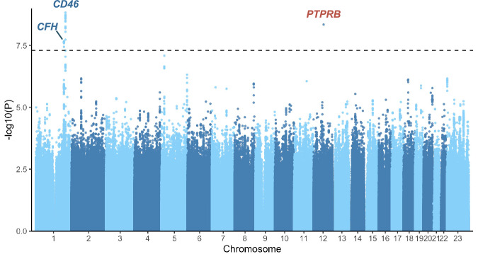
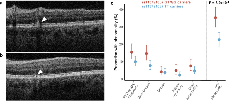
Central serous chorioretinopathy is an eye disease characterized by fluid buildup under the central retina whose etiology is not well understood. Abnormal choroidal veins in central serous chorioretinopathy patients have been shown to have similarities with varicose veins. To identify potential mechanisms, we analyzed genotype data from 1,477 patients and 455,449 controls in FinnGen. We identified an association for a low-frequency (allele frequency = 0.5%) missense variant (rs113791087) in PTPRB, the gene encoding vascular endothelial protein tyrosine phosphatase (odds ratio=2.85, P = 4.5 × 10⁻⁹). This was confirmed in a meta-analysis of 2,452 patients and 865,767 controls from 4 studies (odds ratio=3.06, P = 7.4 × 10⁻¹⁵). Rs113791087 was associated with a 56% higher prevalence of retinal abnormalities (35.3% vs 22.6%, P = 8.0 × 10⁻⁴) in 708 UK Biobank participants and, surprisingly, with increased risk of varicose veins (odds ratio=1.31, P = 2.3 × 10⁻¹¹) and reduced risk of glaucoma (odds ratio=0.82, P = 6.9 × 10⁻⁹). Predicted loss-of-function variants in PTPRB, though rare in number, were associated with central serous chorioretinopathy in All of Us (odds ratio=17.09, P = 0.018). These findings highlight the significance of vascular endothelial protein tyrosine phosphatase in diverse ocular and systemic veno-vascular diseases.


Conflict of interest statement

Competing interests: Dr. Rossin and Dr. Rämö are named inventors on a provisional patent application that describes the secondary use of intravitreal Anti-Ang2 medications for use in the treatment of central serous chorioretinopathy. Dr. Ellinor receives sponsored research support from Bayer AG, IBM Research, Bristol Myers Squibb, Pfizer and Novo Nordisk; he has also served on advisory boards or consulted for MyoKardia and Bayer AG. The remaining authors declare no competing interests.

Figures

Fig. 1
Fig. 1. Genome-wide association study of central serous chorioretinopathy in FinnGen.
A genome-wide association study of central serous chorioretinopathy was conducted, including 1477 patients with central serous chorioretinopathy and 455,449 controls from the FinnGen study. Each genomic variant is plotted as a data point, with P-values derived using logistic regression as implemented in Regenie (v2.2.4) shown on the y-axis on a logarithmic scale and chromosomal position shown on the x-axis. All P values are two-sided and were not adjusted for multiple comparisons. The genome-wide significance threshold (P = 5×10⁻⁸) is shown with a dashed line. In each of three loci reaching genome-wide significance, the nearest protein-coding gene to the lead variant is labeled (blue = previously reported loci, red = novel locus).
Fig. 2
Fig. 2. Associations of the PTPRB missense variant rs113791087 with central serous chorioretinopathy in 4 studies.
The association of rs113791087 with central serous chorioretinopathy (CSC) was examined in 4 different studies. In all biobank-based studies (FinnGen, Million Veteran Program [MVP] and All of Us) included in the meta-analysis, patients with CSC were identified based on International Statistical Classification of Diseases codes, and all participants with age-related macular degeneration were excluded from patients and controls following a harmonized study protocol. In the chronic CSC (cCSC) cohort, patients were identified from ophthalmological clinics based on expert review. Odds ratios (OR) and P-values were derived using logistic regression as implemented in Regenie (v2.2.4) in FinnGen, SAIGE (v1.3.0) in MVP, and Regenie (v3.2.2) in All Of Us. Association estimates and two-sided p-values were previously derived in the European cCSC cohort using the firth bias-corrected likelihood ratio test. An inverse-weighted fixed-effects meta-analysis was also conducted to combine data from all studies. The point estimates of the odds ratios are shown with circles for the individual studies and with a square for the meta-analysis, and 95% confidence intervals (CI) are denoted with lines. For the All of Us cohort, the upper range of the confidence interval is truncated (arrow). We observed no statistically significant heterogeneity for rs113791087 in the meta-analysis (I2 = 0.48, Q-statistic = 5.8, Q-statistic P value = 0.12). All P-values are two-sided and were not adjusted for multiple comparisons.
Fig. 3
Fig. 3. Retinal abnormalities in optical coherence tomography images by PTPRB rs113791087 genotype status in UK Biobank.
Optical coherence tomography (OCT) images of 266 participants with the rs113791087 G allele (GG or GT genotype) and 442 age-matched participants lacking the G allele (TT genotype) were obtained from UKB. Images were independently evaluated by 3 retina specialists who were blinded to genotype. Each grader was tasked with identifying and categorizing retinal pigment epithelium (RPE) abnormalities according to the following categories: rare drusen (1−5), drusen (>5), pattern dystrophy, pigment epithelial detachment (PED) or nonspecific RPE irregularity, and other rare findings (subretinal fluid, pachychoroid pigment epitheliopathy, atrophy, intraretinal fluid, or evidence of central serous chorioretinopathy). Panel a depicts a B-scan superior to the fovea showing an area of RPE irregularity and pigment migration (arrowhead) from a randomly selected participant, who had the TT genotype. Panel b depicts a B-scan inferior to the fovea showing a second area of RPE irregularity and pigment migration (arrowhead) in the same participant. Panel c shows the prevalence of abnormalities (denoted with circles) for participants with the GG or GT genotype (red) and TT genotype (blue). Statistical significance was evaluated using logistic regression, including age, sex, examiner and the first 10 PCs as covariates. The P-value is two-sided and not adjusted for multiple comparisons. Binomial proportion 95% confidence intervals were calculated using the Agresti-Coull method. Optical coherence tomography images are reproduced by kind permission from UK Biobank ©.
Fig. 4
Fig. 4. Phenome-wide association study of the PTPRB missense variant rs113791087 in FinnGen.
To identify potential pleiotropic associations of the rs113791087 variant, a phenome-wide association study was conducted including 2,469 phenotypes as outcomes using logistic regression as implemented in Regenie (v2.2.4). All P values are two-sided and were not adjusted for multiple comparisons. Data are shown for all phenotypes that were at least nominally associated (P < 0.05) with rs113791087. The negative common logarithm of each P value is shown on the y-axis. The genome-wide significance threshold (P = 5×10⁻⁸) is shown with a dashed line. The direction of association with the risk of each disease is denoted by symbols (arrow up = increased risk, arrow down = decreased risk).
Fig. 5
Fig. 5. Cross-study meta-analyses of other diseases most significantly associated with the PTPRB missense variant rs113791087.
The distinct diseases most significantly associated with rs113791087 in the phenome-wide association study of rs113791087 in FinnGen were carried forward for multi-study meta-analyses, including available data from FinnGen, UK Biobank (UKB), Million Veteran Program (MVP), and All of Us (AoU). Pulmonary embolism and deep vein thrombosis were examined separately as the major subtypes of venous thromboembolism, and primary open-angle glaucoma was examined separately as a major subtype of glaucoma. Odds ratios (OR) and P values were derived using logistic regression as implemented in Regenie (v2.2.4) in FinnGen, SAIGE (v1.3.0) in MVP, and Regenie (v3.2.2) in All Of Us. Association results from contributing studies were combined in an inverse variance-weighted meta-analysis. Point estimates of the odds ratios are shown with circles for each phenotype, and 95% confidence intervals (CI) are denoted with lines. All P-values are two-sided and were not adjusted for multiple comparisons.
Fig. 6
Fig. 6. Disease associations of predicted loss-of-function variants in PTPRB among UK Biobank and All of Us participants.
The risk of ocular and vascular diseases of interest was evaluated for participants with a predicted loss-of-function (pLOF) variant in PTPRB in All of Us and UK Biobank using logistic regression among unrelated individuals. Due to lack of ophthalmological outpatient clinic data in UK Biobank participants, patients with central serous chorioretinopathy were only evaluated in All of Us. Examined diseases were selected based on the most significant associations observed for rs113791087 in FinnGen. Pulmonary embolism and deep vein thrombosis were examined separately as the major subtypes of venous thromboembolism, and primary open-angle glaucoma was examined separately as a major subtype of glaucoma. Association results from UK Biobank and All of Us were combined in an inverse variance-weighted meta-analysis. Point estimates of the odds ratios (OR) are shown with circles for each phenotype, and 95% confidence intervals (CI) are denoted with lines. All P-values are two-sided and were not adjusted for multiple comparisons.

Update of

References

    1. Kitzmann, A. S., Pulido, J. S., Diehl, N. N., Hodge, D. O. & Burke, J. P. The Incidence of Central Serous Chorioretinopathy in Olmsted County, Minnesota, 1980–2002. Ophthalmology115, 169–173 (2008). - PubMed
    1. Feenstra H. M. A. et al. Central serous chorioretinopathy: An evidence-based treatment guideline. Prog Retin Eye Res. 101236 (2024). - PubMed
    1. van Rijssen, T. J. et al. Central serous chorioretinopathy: Towards an evidence-based treatment guideline. Prog. Retin Eye Res73, 100770 (2019). - PubMed
    1. Sahoo, N. K. et al. Prevalence and Profile of Central Serous Chorioretinopathy in an Indian Cohort. Nepal J. Ophthalmol.11, 5–10 (2019). - PubMed
    1. Montero, J. A. Ruiz-Moreno JM. Optical coherence tomography characterisation of idiopathic central serous chorioretinopathy. Br. J. Ophthalmol.89, 562–564 (2005). - PMC - PubMed

Publication types

Substances

LinkOut - more resources