. 2025 May 3;13(1):90.
doi: 10.1186/s40478-025-01998-x.

Systemic IGF-1 administration prevents traumatic brain injury induced gut permeability, dysmorphia, dysbiosis, and the increased number of immature dentate granule cells

Affiliations

Systemic IGF-1 administration prevents traumatic brain injury induced gut permeability, dysmorphia, dysbiosis, and the increased number of immature dentate granule cells

Lavanya Venkatasamy et al. Acta Neuropathol Commun. .

Abstract

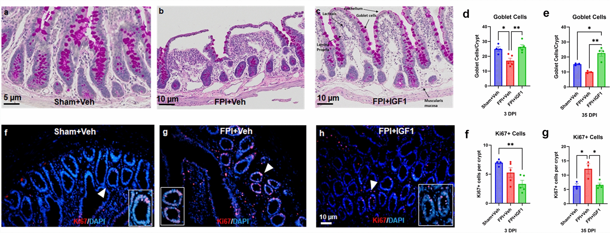
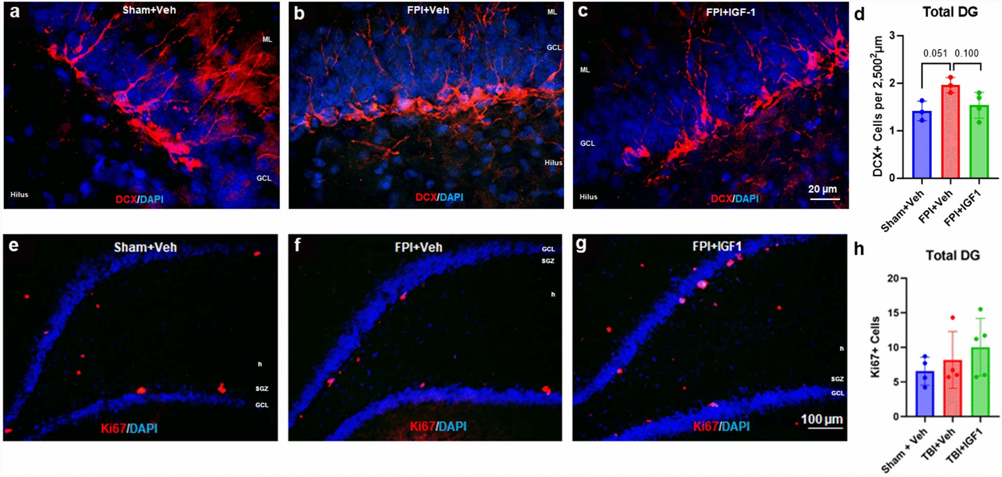
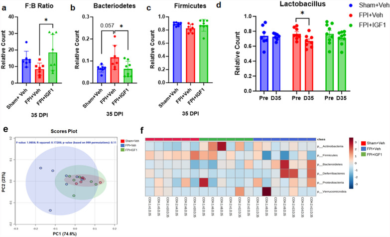
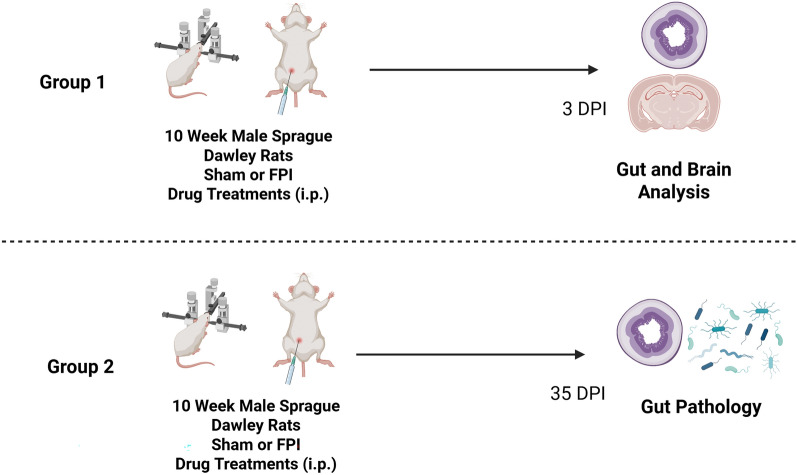
Traumatic brain injury (TBI) occurs in 2-3 million Americans each year and is a leading cause of death and disability. Among the many physiological consequences of TBI, the hypothalamic pituitary axis (HPA) is particularly vulnerable, including a reduction in growth hormone (GH) and insulin-like growth factor (IGF-1). Clinical and preclinical supplementation of IGF-1 after TBI has exhibited beneficial effects. IGF-1 receptors are prominently observed in many tissues, including in the brain and in the gastrointestinal (GI) system. In addition to causing damage in the brain, TBI also induces GI system damage, including inflammation and alterations to intestinal permeability and the gut microbiome. The goal of this study was to assess the effects of systemic IGF-1 treatment in a rat model of TBI on GI outcomes. Because GI dysfunction has been linked to hippocampal dysfunction, we also examined proliferation and immature granule cells in the hippocampal dentate gyrus. 10-week-old male rats were treated with an intraperitoneal (i.p.) dose of IGF-1 at 4 and 24 h after lateral fluid percussion injury (FPI). At 3- and 35-days post-injury (DPI), gut permeability, gut dysmorphia, the fecal microbiome, and the hippocampus were assessed. FPI-induced permeability of the blood-gut-barrier, as measured by elevated gut metabolites in the blood, and this was prevented by the IGF-1 treatment. Gut dysmorphia and alterations to the microbiome were also observed after FPI and these effects were ameliorated by IGF-1, as was the increase in immature granule cells in the hippocampus. These findings suggest that IGF-1 can target gut dysfunction and damage after TBI, in addition to its role in influencing adult hippocampal neurogenesis.

Keywords: Dentate gyrus; Fluid percussion injury (FPI); Gastrointestinal system; Growth hormone; Gut microbiome; Hippocampus; Metabolite; Neurogenesis; Newborn neurons; TBI.


Conflict of interest statement

Declarations. Ethics approval and consent to participate: All the animal experimental protocols were approved by the Institutional Animal Care Committee (IACUC) of Texas A&M Health Science Center (AUP #2010-0140). Consent for publication: Not applicable. Competing interests: The authors declare no competing interests.

Figures

Fig. 1
Fig. 1
IGF-1 treatment rescues early FPI-induced gut permeability. Blood-gut barrier permeability was assessed at 3 DPI by measuring serum levels of LPS, iFABP and Muc-2 [–80]. In a, there was a significant increase in the serum concentration of LPS in the FPI + Veh rats (p < 0.0001 vs. Sham + Veh) that was significantly ameliorated by IGF-1 treatment (p < 0.001 vs. TBI + Veh). In b, levels of serum iFABP were significantly elevated in FPI + Veh as compared to the Sham + Veh rats (p < 0.0001), and this increase was significantly attenuated in the FPI + IGF-1 rats (p < 0.001 vs. FPI + Veh). In c, the concentration of Muc-2 (520 kD) was significantly elevated in FPI + Veh rats compared to the Sham + Veh rats (p < 0.0001) and reduced in FPI + IGF-1 rats (p < 0.001 vs. FPI + Veh). These data suggest that the blood-gut barrier and the mucus barrier may deteriorate after FPI and this is prevented by IGF-1. Data are represented as Mean ± SEM; n = 8–10 per group. Note, 3 rats in the FPI + IGF-1 group were omitted from Mucin-2 analysis due to limited serum availability. ***p < 0.001; ****p < 0.0001
Fig. 2
Fig. 2
IGF-1 treatment reduces FPI-induced chronic gut dysmorphia. To assess gut morphology, villi and crypts were analyzed in the ileum using H&E staining at 3 (af) and 35 (jo) DPI. Note the normal appearance of the villi and crypts in sham rats (black arrowhead; a,b). Also note that at the 3 DPI timepoint, the villi are beginning to show a rounded stumpy appearance (green arrowhead; c,d), but the quantitative results are not significant at this time point (g, h). These morphological differences evolve over time so that they appear more pronounced at 35 DPI, such that they have a significantly reduced length:width ratio (p < 0.01 vs. Sham + Veh), and this was significantly increased by IGF-1 treatment (p < 0.01) (o). Importantly, the villi from the IGF-1 treated rats appears much more sham-like (e,f). In p, the number of crypts per villus in the ileum was increased after FPI (p < 0.05 vs. Sham + Veh), and IGF-1 treatment after FPI significantly reduced this effect (p < 0.05). For all rats, 3 slides were assessed, with 3 sections per slide measured. Scale bar in e,n = 10 µm for A,C,E,J,L,N; scale bar in f,o = 5 µm for B,D,F,L,M,O. Data are represented as Mean ± SEM. N for 3 DPI: 4 Sham + Veh, 5 FPI + Veh, 5 FPI + IGF1. N for 35 DPI: 3 Sham + Veh, 3 FPI + Veh, 4 FPI + IGF1. *p < 0.05; **p < 0.01
Fig. 3
Fig. 3
Influence of FPI and IGF-1 on goblet cells and intestinal epithelial stem cells (IESC). PAS staining was used to assess the number of goblet cells per crypt in the ileum at 3 (ac) and 35 DPI. Quantitative analysis revealed that FPI significantly reduced the number of goblet cells per crypt at 3 DPI (d) and 35 DPI (e) and this was increased by IGF-1 treatment. Proliferating IESCs in the crypts were determined by quantifying Ki67 + labeled cells at 3 (i) and 35 (j) DPI. In f–h, representative micrographs of Ki67 + staining in the crypts of the ileum at 35 DPI, with areas designated by the white arrow enlarged in the corresponding inset. At 3 DPI (i), the FPI + Veh showed a non-significant reduction (p = 0.2152, NS) in the number of Ki67 + cells in the crypts. The FPI + IGF-1 group exhibited a significant reduction (p < 0.01 vs. to Sham + Veh) in the Ki67 + cells in the crypts. Conversely at 35 DPI (j), FPI significantly increased the number of Ki67 + cells, and this was significantly reduced by IGF-1 treatment. This suggests that FPI resulted in a delayed increase in IESC proliferation, whereas IGF-1 reduced the number of Ki67 + cells at both timepoints examined. For all rats, 3 slides were assessed, with 3 sections per slide measured. Data are represented as Mean ± SEM; N for 3 DPI: 4 Sham + Veh, 5 FPI + Veh, 5 FPI + IGF1. N for 35 DPI: 3 Sham + Veh, 3 FPI + Veh, 4 FPI + IGF1. *p < 0.05; **p < 0.01. Scale bar in a = 5 µm; scale bar in b,c = 10 µm; scale bar in h = 10 µm for f–h
Fig. 4
Fig. 4
IGF-1 ameliorates the FPI-induced increase in immature neurons in the adult hippocampus. Immature neurons in the hippocampal dentate gyrus were assessed by stereological quantification of the number of DCX-labeled cells at 3 DPI (ac). The results show that FPI increased the number of DCX + cells (p = 0.051), and this was mitigated by IGF-1 treatment (d). The number of proliferative cells in the dentate gyrus (DG) was assessed by quantifying the number of Ki67-labeled cells (eg) in the hilus (h), granule cell layer (GCL), and subgranular zone (SGZ). No significant differences in the number of Ki67 + cells in the hippocampus were identified at 3 DPI (h). Scale bar in c = 20 µm for (ac). Data are represented as Mean ± SEM; n = 3–4 per group
Fig. 5
Fig. 5
Gut dysbiosis after FPI is improved by IGF-1. Gut bacterial composition was assessed from fecal samples at 35 DPI. The ratio of Firmicutes to Bacteroidetes (F:B), an estimate of gut health, was significantly increased in the FPI + IGF-1 treated rats compared to the FPI + Veh rats (p < 0.05) (a). The relative count of Bacteriodetes was increased in FPI + Veh rats (p = 0.057, NS vs. Sham + Veh), and this was significantly reduced with IGF-1 treatment (p < 0.05 vs. FPI + Veh) (b). There were no significant changes identified in Firmicutes (c). In d, the abundance of Lactobacillus was significantly decreased in the FPI + Veh rats at 35 DPI compared to baseline (p < 0.05), while there were no significant differences identified at 35 DPI in either the Sham + Veh or FPI + IGF-1 groups. In e, principal component analysis (PCA) using the partial Least-Squares Discriminant Analysis (PLS-DA shows that the major phyla for Sham + Veh and FPI + IGF-1 rats cluster more closely together than do the FPI + Veh. In f, heat map expression levels further demonstrate that FPI + Veh rats have an altered gut bacterial repertoire, compared to Sham + Veh, and that the FPI + IGF-1 rats are more similar to sham rats. Data are represented as Mean ± SEM; n = 4–5 per group. *p < 0.05; **p < 0.01
Fig. 6
Fig. 6
Experimental Design for the study. Schematic diagram depicting the experimental design showing that rats received either sham or FPI at 10-weeks-of age, followed by IGF-1 or vehicle at 4 and 24 hours after surgery. Tissue was then collected for analysis at either 3 or 35 days post injury (DPI)

Similar articles

References

    1. Maas AIR et al (2017) Traumatic brain injury: integrated approaches to improve prevention, clinical care, and research. Lancet Neurol 16(12):987–1048 - PubMed
    1. Coronado VG et al (2012) Trends in traumatic brain injury in the U.S. and the public health response: 1995–2009. J Saf Res 43(4):299–307 - PubMed
    1. Ahmed S et al (2017) Traumatic brain injury and neuropsychiatric complications. Indian J Psychol Med 39(2):114–121 - PMC - PubMed
    1. Collaborators GUND (2021) Burden of neurological disorders across the US from 1990–2017: a global burden of disease study. JAMA Neurol 78(2):165–176 - PMC - PubMed
    1. Elser H et al (2023) Head injury and long-term mortality risk in community-dwelling adults. JAMA Neurol 80(3):260–269 - PMC - PubMed

Publication types

MeSH terms

Substances