. 2025 May 3;24(1):132.
doi: 10.1186/s12943-025-02324-8.

Lymph node macrophage-targeted interferon alpha boosts anticancer immune responses by regulating CD169-positive phenotype of macrophages

Affiliations

Lymph node macrophage-targeted interferon alpha boosts anticancer immune responses by regulating CD169-positive phenotype of macrophages

Ryo Fukuda et al. Mol Cancer. .

Abstract

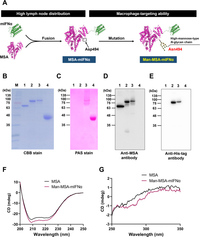
Background: CD169+ macrophages in lymph nodes (LNs) activate cytotoxic T lymphocytes (CTLs), which play a crucial role in anticancer immunity, through antigen presentation and co-stimulation by CD169. Interferon alpha (IFNα) is capable of inducing the CD169+ phenotype of macrophages; however, its clinical applications have been hindered by pharmacokinetic limitations-low LN distribution and an inability to target macrophages. To overcome these issues, this study genetically fused mouse IFNα (mIFNα) with mannosylated mouse serum albumin (Man-MSA), and investigated the antitumor effects of the hybrid protein (Man-MSA-mIFNα) or its add-on effects with programmed death-ligand 1 (PD-L1) blockade.

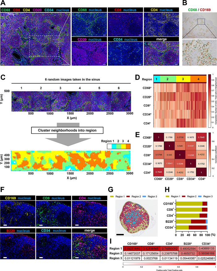
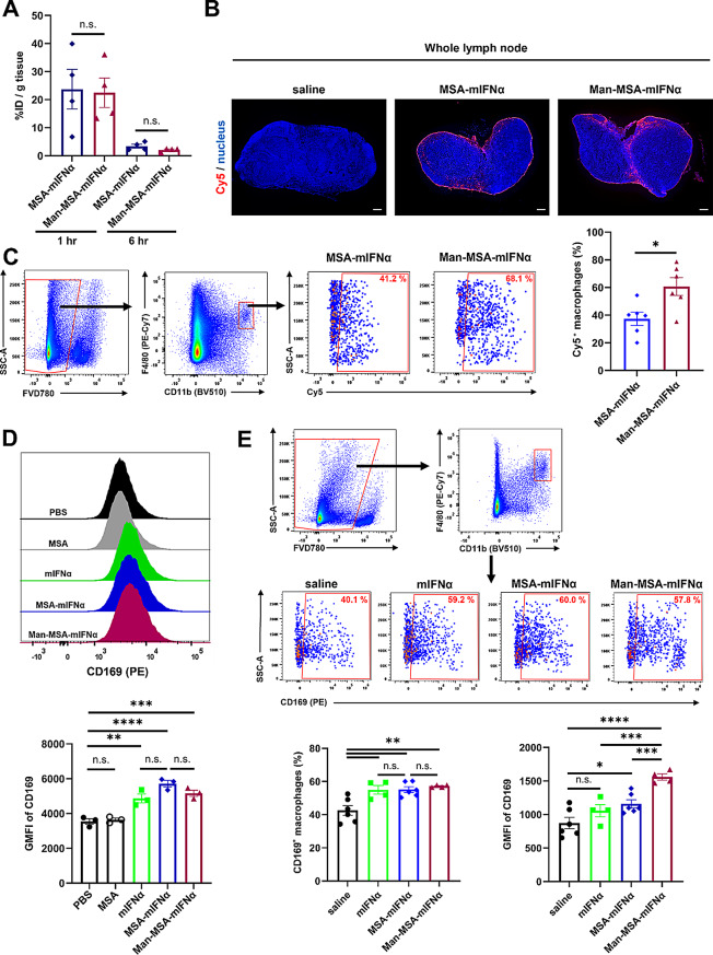
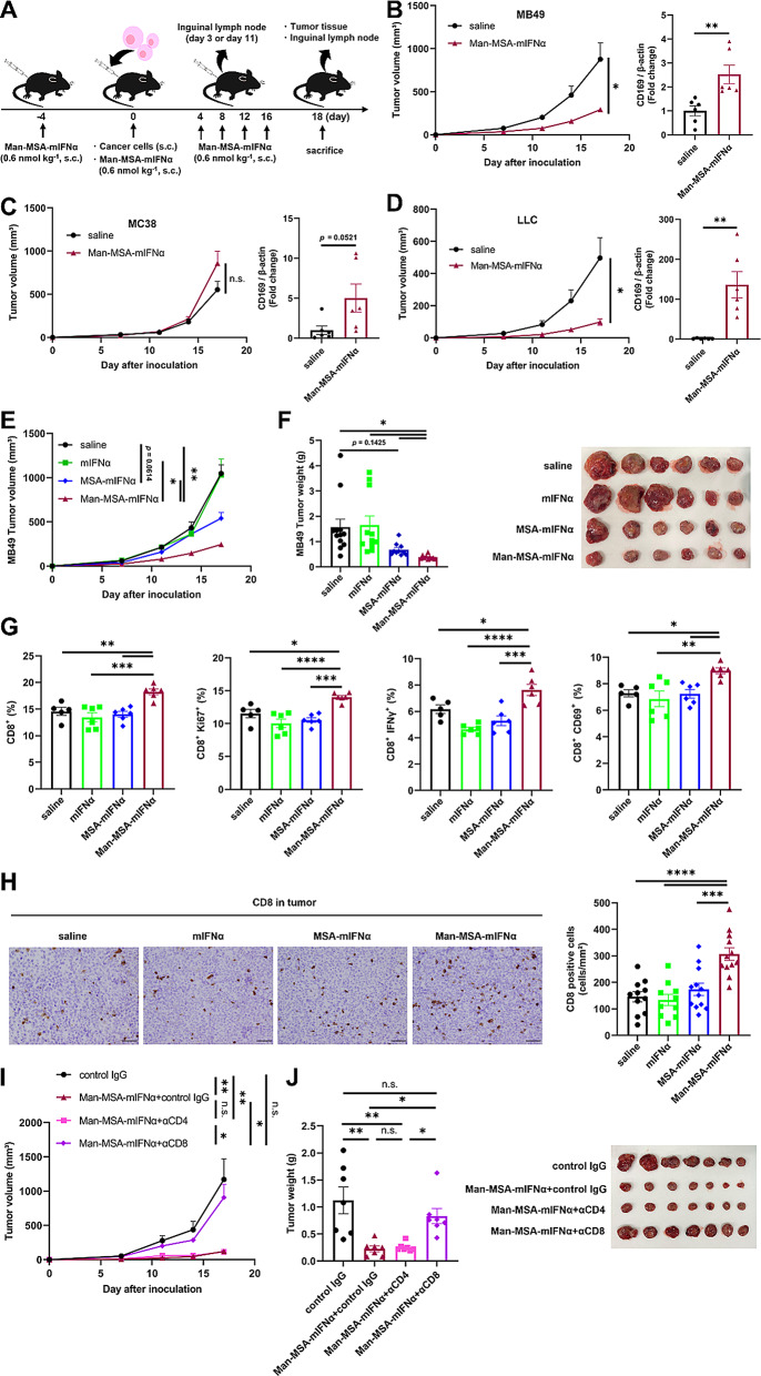
Methods: To confirm the possibility of CD169+ macrophage-mediated T cell priming, positional information about individual immune cells in LNs of cancer patients was obtained. Traits of Man-MSA-mIFNα, which was prepared using Pichia pastoris to form the high-mannose structure, were characterized by several physicochemical methods. To evaluate the lymphatic drainage of Man-MSA-mIFNα, radioiodine or Cy5-labeled Man-MSA-mIFNα was subcutaneously administered in mice, and then the radioactivity or fluorescence in LNs was analyzed. CD169-diphtheria toxin (DT) receptor (CD169-DTR) mice in which LN CD169+ macrophages can be depleted by DT injection were used to verify whether the antitumor effect of Man-MSA-mIFNα is dependent on LN CD169+ macrophages.

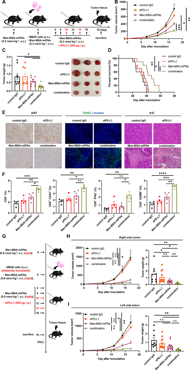
Results: Multiplex tissue imaging predicted close proximity of CD169+ macrophages and T cells and positive correlation between the number of CD169+ macrophages and T cells in neighborhoods in LNs of cancer patients. Physicochemical analyses showed that Man-MSA-mIFNα was formed from the fusion of the intact Man-MSA and mIFNα. Man-MSA-mIFNα efficiently induced the CD169+ phenotype of macrophages by its high LN distribution and macrophage-targeting capability, and significantly exerted antitumor activity through CD8+ T cell activation in the LNs, whereas its antitumor effects were canceled in CD169-DTR mice. Finally, combination therapy with PD-L1 blockade markedly suppressed tumor growth in MB49-bearing mice, which exhibit resistance to PD-L1 blockade monotherapy.

Conclusions: The present study successfully designed and developed Man-MSA-mIFNα, which efficiently induces the CD169+ phenotype in LN macrophages, contributing to the antitumor immunity. The findings suggest that our novel strategy targeting CD169⁺ macrophages could be a promising immunotherapy for cancer patients who are unresponsive to immune checkpoint inhibitors.

Keywords: Albumin; CD169; Cancer immunotherapy; Drug delivery system; IFNα; Lymph node; Macrophage; Targeted therapy.


Conflict of interest statement

Declarations. Ethics approval and consent to participate: The study design was approved by the Institutional Review Board of Kumamoto University (#1169) in accordance with the guidelines for Good Clinical Practice and the Declaration of Helsinki. Paraffin-embedded LN samples were prepared from specimens obtained from patients diagnosed with colorectal cancer that were surgically resected at Izumi General Hospital (Izumi, Kagoshima, Japan). Written informed consent was obtained from all patients, and the study design was approved by the review board (#57). The need for individual patient consent for inclusion in the study was waived by the Institutional Review Board of Kumamoto University (#1169). All animal experiments were conducted using procedures approved by the experimental animal ethics committee at Kumamoto University (A2021-021). Consent for publication: Not applicable. Competing interests: The authors declare no competing interests.

Figures

Fig. 1
Fig. 1
Spatial analysis of regional LN cells in cancer patients and tumor-bearing mice. A) Multiple immunostaining for regional LNs of colorectal cancer patients. Scale bar, left: 200 μm, magnified images: 50 μm. B) Regional LNs of colorectal cancer patients were stained with CD68 (green) and CD169 (brown). The representative sinus area in the LN (upper, scale bar: 200 μm) was magnified (lower, scale bar: 50 μm). C) Representative 2D (X, Y) tissue images (500 × 500 μm) in the sinus (upper) and positional plot of the neighborhoods color coded by the region type (lower). D) Heatmap of the neighborhood composition (the number of cells per total cells in neighborhood). E) Heatmap of the Pearson correlation coefficients of the number of cells per neighborhood from all regions. F) Multiple immunostaining for regional LNs of MB49-bearing mice. Scale bar: 50 μm. G) Positional plot of the neighborhoods color coded by the region type. Scale bar: 500 μm. H) Percentage of each cell in each region. I) Heatmap of the number of positive cells in each region per total positive cells in all area. These results are representative of two independent experiments
Fig. 2
Fig. 2
Physicochemical characterization of Man-MSA-mIFNα. A) Experimental scheme for the development of Man-MSA-mIFNα. B) CBB stain after SDS-PAGE. C) PAS stain after SDS-PAGE. D, E) MSA and His-tag were detected by western blot analysis. M, Molecular weight marker; 1, MSA; 2, MSA-mIFNα; 3, Man-MSA-mIFNα; 4, AGP. F) Far-UV and G) near-UV spectra were recorded from 200 to 250 nm and from 250 to 350 nm, respectively. These results are representative of three independent experiments
Fig. 3
Fig. 3
Intralymph node distribution of Man-MSA-mIFNα and its capacity to induce CD169+ phenotype of macrophages. A) LN distribution was measured 1 h and 6 h after administration (s.c.) of each protein (n = 4). This experiment was performed once. B) Distribution of Cy5-labeled fusion proteins in LNs observed 1 h after subcutaneous administration. Scale bar: 200 μm. C) The rate of Cy5-incorporated cells in CD11b+ F4/80+ macrophages 1 h after subcutaneous administration was evaluated by flow cytometry (n = 6). D) J774.1 cells were incubated with each protein, followed by the evaluation of CD169 expression by flow cytometry (n = 3). A representative graph (upper) and geometric mean fluorescence intensity (GMFI) of CD169 (lower). E) Seventy-two hours after subcutaneous administration, inguinal LNs were harvested, and CD169 expression in CD11b+ F4/80+ cells was evaluated by flow cytometry (n = 4–6). The titer of mIFNα in each administration group was set to be equal by performing an in vitro experiment using J774.1 cells. A representative gating strategy for CD11b+ F4/80+ CD169+ cells (upper), CD169-positive macrophages (%) in CD11b+ F4/80+ cells (bottom left) and GMFI of CD169 in CD11b+ F4/80+ cells (bottom right). These results (B-E) are representative of two independent experiments. Data are averages ± S.E. *p < 0.05, **p < 0.01, ***p < 0.001, ****p < 0.0001, n.s.=nonsignificant; BV510 = Brilliant Violet 510; FVD780 = Fixable Viability Dye eFluor 780; PE = phycoerythrin; SSC-A = side scatter-peak area
Fig. 4
Fig. 4
The effect of Man-MSA-mIFNα on tumor growth and CD8+ T cell activation in tumor-bearing mice. A) The experimental protocol for the evaluation of the antitumor effect of Man-MSA-mIFNα in tumor-bearing mice. B) Subcutaneous tumor volume was measured in MB49-bearing mice at each time point (n = 5–6). Inguinal LNs were harvested from MB49-bearing mice on the 3rd day after tumor inoculation and analyzed for CD169 expression by western blotting (n = 6). C) Subcutaneous tumor volume was measured in MC38-bearing mice at each time point (n = 6). Inguinal LNs were harvested from MC38-bearing mice on the 3rd day after tumor inoculation and analyzed for CD169 expression by western blotting (n = 6). D) Subcutaneous tumor volume was measured in LLC-bearing mice at each time point (n = 6). Inguinal LNs were harvested from LLC-bearing mice on the 11th day after tumor inoculation and analyzed for CD169 expression by western blotting (n = 6). These results (B-D) are representative of two independent experiments. E) Subcutaneous tumor volume was measured in MB49-bearing mice at each time point (n = 10–12). F) Subcutaneous tumor weight (n = 10–12) (left) and the representative image of tumors (right) on the 18th day after tumor inoculation. These results (E, F) represent combined data of two independent experiments. G) Inguinal LNs were harvested from MB49-bearing mice on the 18th day after tumor inoculation, followed by the evaluation of the percentage of CD8+ cells, Ki67+ CD8+ cells, IFNγ+ CD8+ cells, and CD69+ CD8+ cells in live cells by flow cytometry (n = 5–6). These results are representative of two independent experiments. H) Subcutaneous tumors on the 18th day after tumor inoculation were evaluated for tumor infiltration of CD8+ cells by immunohistochemistry (n = 10–12). Representative images (left) and quantification (right) of CD8+ cells in tumors. This result represents combined data of two independent experiments. Scale bar: 50 μm. I) Subcutaneous tumor volume was measured in MB49-bearing mice at each time point (n = 7). J) Subcutaneous tumor weight (n = 7) (left) and the representative image of tumors (right) on the 18th day after tumor inoculation. (I, J) MB49-bearing mice were depleted of CD4+ or CD8+ T cells and then received Man-MSA-mIFNα treatment. These results (I, J) are representative of two independent experiments. Data are averages ± S.E. *p < 0.05, **p < 0.01, ***p < 0.001, ****p < 0.0001, n.s.=nonsignificant
Fig. 5
Fig. 5
The effect of Man-MSA-mIFNα on tumor growth and CD8+ T cell activation in CD169++ macrophage-depleted mice. A) The experimental protocol for the evaluation of the antitumor effect of Man-MSA-mIFNα in CD169+ macrophage-depleted MB49-bearing mice. B) Inguinal LNs were harvested from MB49-bearing WT mice or MB49-bearing CD169-DTR mice on the 18th day after tumor inoculation, followed by the evaluation of the percentage of CD169+ cells in CD11b+ F4/80+ cells by flow cytometry (n = 6). C) Subcutaneous tumor volume was measured in MB49-bearing mice at each time point (n = 5–6). D) Subcutaneous tumor weight (n = 5–6) (left) and the representative image of tumors (right) on the 18th day after tumor inoculation. E) Inguinal LNs were harvested from MB49-bearing mice on the 18th day after tumor inoculation, followed by the evaluation of the percentage of CD8+ cells, Ki67+ CD8+ cells, IFNγ+ CD8+ cells, and CD69+ CD8+ cells in live cells by flow cytometry (n = 6). F) Subcutaneous tumors on the 18th day after tumor inoculation were evaluated for tumor infiltration of CD8+ cells by immunohistochemistry (n = 5–6). Representative images (left) and quantification (right) of CD8+ cells in tumors. Scale bar: 50 μm. These results are representative of two independent experiments. Data are averages ± S.E. ****p < 0.0001, n.s.= nonsignificant
Fig. 6
Fig. 6
The effect of combination therapy on tumor growth and CD8+T cell activation in tumor-bearing mice. A) The experimental protocol for the evaluation of the antitumor effect of combination therapy in MB49-bearing mice. B) Subcutaneous tumor volume was measured in MB49-bearing mice at each time point (n = 8–9). C) Subcutaneous tumor weight (n = 8–9) (left) and the representative image of tumors (right) on the 18th day after tumor inoculation. These results (B, C) represent combined data of two independent experiments. D) Survival time was observed in MB49-bearing mice treated by PD-L1 blockade and/or Man-MSA-mIFNα (n = 10). This result is representative of two independent experiments. E) Representative images of immunostaining for Ki67, terminal deoxynucleotidyl transferase dUTP nick end labeling (TUNEL), and H.E. staining of tumors from MB49-bearing mice on the 18th day after tumor inoculation. Scale bar: 50 μm. F) Subcutaneous tumors were harvested from MB49-bearing mice on the 18th day after tumor inoculation, followed by the evaluation of the percentage of CD8+ cells, CD44+ CD8+ cells, IFNγ+ CD8+ cells, and GZMB+ CD8+ cells in live cells by flow cytometry (n = 4). These results (E, F) are representative of two independent experiments. G) The experimental protocol for the evaluation of the antitumor effect in bilateral MB49-bearing mice. Subcutaneous tumor volume and weight were measured in MB49-bearing mice at both the H) local tumor (right side tumor) and the I) distant tumor (left side tumor) (n = 9–12). These results (H, I) represent combined data of two independent experiments. Data are averages ± S.E. *p < 0.05, **p < 0.01, ***p < 0.001, ****p < 0.0001, n.s.= nonsignificant; αPD-L1 = alpha programmed death-ligand 1

References

    1. Yang Y, Attwood K, Bshara W, Mohler JL, Guru K, Xu B, Kalinski P, Chatta G. High intratumoral CD8(+) T-cell infiltration is associated with improved survival in prostate cancer patients undergoing radical prostatectomy. Prostate. 2021;81:20–8. - PMC - PubMed
    1. Ribas A, Wolchok JD. Cancer immunotherapy using checkpoint Blockade. Science. 2018;359:1350–5. - PMC - PubMed
    1. Mellman I, Chen DS, Powles T, Turley SJ. The cancer-immunity cycle: indication, genotype, and immunotype. Immunity. 2023;56:2188–205. - PubMed
    1. Asano K, Nabeyama A, Miyake Y, Qiu CH, Kurita A, Tomura M, Kanagawa O, Fujii S, Tanaka M. CD169-positive macrophages dominate antitumor immunity by crosspresenting dead cell-associated antigens. Immunity. 2011;34:85–95. - PubMed
    1. Zhang Y, Li JQ, Jiang ZZ, Li L, Wu Y, Zheng L. CD169 identifies an anti-tumour macrophage subpopulation in human hepatocellular carcinoma. J Pathol. 2016;239:231–41. - PubMed

MeSH terms