

Enhanced Bone Regeneration Using Demineralized Dentin Matrix: A Comparative Study in Alveolar Bone Repair
- PMID: 40319772
- PMCID: PMC12124616
- DOI: 10.1016/j.identj.2025.03.026
Enhanced Bone Regeneration Using Demineralized Dentin Matrix: A Comparative Study in Alveolar Bone Repair
Abstract
Objectives: Alveolar bone resorption following tooth extraction presents significant challenges for implant-supported rehabilitations. Demineralised dentin matrix (DDM) has emerged as a promising scaffold for bone tissue regeneration. This study evaluates the bone-regenerating potential of varying degrees of dentin demineralisation.
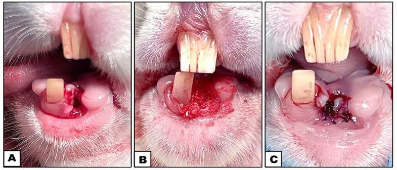
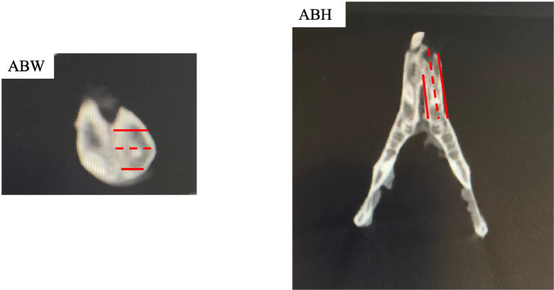
Materials and methods: Thirty-two male white New Zealand rabbits underwent extraction of the left mandibular anterior tooth and were assigned to 4 groups: undemineralised dentin matrix (UDDM), partially demineralised dentin matrix (PDDM), completely demineralised dentin matrix (CDDM), and a control group with no treatment. At 4 and 8 weeks post extraction, cone-beam computed tomography (CBCT) was used to assess alveolar bone height and width. Histological analyses using H&E and Masson trichrome stains evaluated new bone formation, and immunohistochemistry detected osteopontin expression.
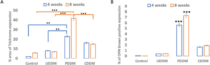
Results: CBCT imaging revealed progressive increases in alveolar bone height and width across all groups over time. Histological analysis showed new bone formation in all groups, with the PDDM group demonstrating closer integration of newly formed bone trabeculae compared with the others. IHC results showed higher osteopontin expression in the PDDM group, highlighting its superior bone-inductive potential.
Conclusion: Among the tested materials, PDDM exhibited the most effective bone induction and tissue regeneration capabilities, outperforming CDDM and UDDM in promoting alveolar bone repair. These findings position PDDM as a valuable scaffold for enhancing bone tissue regeneration in clinical applications.
Clinical relevance: The use of PDDM in tooth extraction sockets significantly promotes efficient and reliable bone regeneration, making it a valuable option for clinical applications in implant dentistry.
Keywords: Alveolar ridge augmentation; Bone regeneration; Cone-beam computed tomography; Demineralised dentin matrix; Immunohistochemistry.
Copyright © 2025 The Authors. Published by Elsevier Inc. All rights reserved.
Conflict of interest statement
Conflict of interest None disclosed.
Figures


Similar articles
-
Evaluation of Effectiveness of Nanocrystalline Hydroxyapatite and Demineralized Bone Matrix Combined with Titanium-platelet Rich Fibrin for Ridge Preservation: A Randomized Controlled Clinical Trial.J Contemp Dent Pract. 2024 Nov 1;25(11):1069-1076. doi: 10.5005/jp-journals-10024-3786. J Contemp Dent Pract. 2024. PMID: 39905614 Clinical Trial.
-
Clinical outcomes of using operating microscope for alveolar ridge preservation: A randomized controlled trial.J Periodontol. 2025 Mar;96(3):230-240. doi: 10.1002/JPER.24-0081. Epub 2024 Oct 15. J Periodontol. 2025. PMID: 39403776 Free PMC article. Clinical Trial.
-
Interventions for replacing missing teeth: alveolar ridge preservation techniques for dental implant site development.Cochrane Database Syst Rev. 2015 May 28;2015(5):CD010176. doi: 10.1002/14651858.CD010176.pub2. Cochrane Database Syst Rev. 2015. Update in: Cochrane Database Syst Rev. 2021 Apr 26;4:CD010176. doi: 10.1002/14651858.CD010176.pub3. PMID: 26020735 Free PMC article. Updated.
-
Does Injectable Platelet-Rich Fibrin Combined With Autogenous Demineralized Dentine Enhance Alveolar Ridge Preservation? A Randomized Controlled Trial.Clin Oral Implants Res. 2025 Feb;36(2):166-177. doi: 10.1111/clr.14372. Epub 2024 Oct 21. Clin Oral Implants Res. 2025. PMID: 39429193 Free PMC article. Clinical Trial.
-
Clinical and histologic outcomes of socket grafting after flapless tooth extraction: a systematic review of randomized controlled clinical trials.J Prosthet Dent. 2015 May;113(5):371-82. doi: 10.1016/j.prosdent.2014.12.009. Epub 2015 Mar 4. J Prosthet Dent. 2015. PMID: 25749077
References
-
- Ten Heggeler J.M., Slot D.E., Weijden G.A. Effect of socket preservation therapies following tooth extraction in non-molar regions in humans: a systematic review. Clin Oral Implants Res. 2011;22:779–788. - PubMed
-
- Diaz C., Del A. TFM; 2016.. Use of Autologous Dental Material as a Graft in the Post-Extraction Socket.
-
- Kim Y.K., Kim S.G., Byeon J.H. Development of a novel bone grafting material using autogenous teeth. Oral Surg Oral Med Oral Pathol Oral Radiol Endod. 2010;109:496.. - PubMed
Publication types
MeSH terms
Substances
LinkOut - more resources
Full Text Sources
Research Materials
