. 2025 May 4;15(1):15597.
doi: 10.1038/s41598-025-99794-z.

Effect of hydrogen sulfide on alpha-synuclein aggregation and cell viability

Affiliations

Effect of hydrogen sulfide on alpha-synuclein aggregation and cell viability

Elena A Ostrakhovitch et al. Sci Rep. .

Abstract

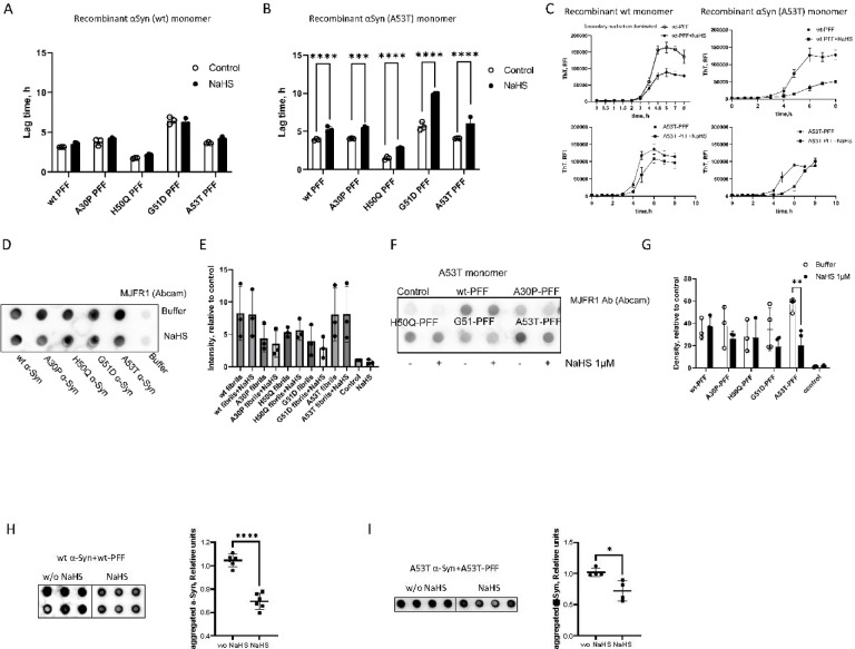
Parkinson's disease (PD) is a progressive neurodegenerative movement disorder characterized by nigrostriatal degeneration and aggregation of α-synuclein (α-Syn) with accumulation of insoluble aggregates in Lewy bodies. Familial mutations in α-Syn are associated with the development of PD. Accumulation of insoluble aggregates results in neuronal toxicity. Identification of compounds that inhibit seeding activity of α-Syn is of great importance. Here we investigate the potential of H2S donor, sodium hydrosulfide (NaHS), to inhibit α-Syn aggregation. We examined the effect of NaHS on fibril growth kinetics and the structural change of α-Syn fibrils formed by self-seeding and cross-seeding of wild-type (wt) and PD familial α-Syn mutations. NaHS slowed both self- and cross-seeded A53T α-Syn fibril formation but not wild-type fibril formation. We observed a decrease in the formed fibril length in vitro. We examined the effect on fibril formation within cells. NaHS significantly reduced the number and filament length of formed oligomers in an α-Syn overexpressing cell model. Furthermore, NaHS rescued viability of A53T α-Syn overexpressing cells seeded with wt- and mutant preformed fibrils. These results support a conformation-specific effect of hydrogen sulfide on alpha-synuclein aggregation and cell viability which deserves further exploration for therapeutic potential.

Keywords: Hydrogen sulfide; Parkinson disease; Protein aggregation; α-Synuclein.


Conflict of interest statement

Declarations. Competing interests: The authors declare no competing interests.

Figures

Fig. 1
Fig. 1
α-Syn familial mutation effects on fibril amplification in vitro and aggregation within cells. (AC) Fibril formation kinetics of human α-Syn monitored by ThT. (A) Comparison of lag time for fibril growth of spontaneous (non-seeded) and (B) self-seeded aggregation of α-Syn monomer. (C) Comparison of lag time for fibril growth of cross-seeded aggregation of α-Syn monomer. Monomeric forms of recombinant wt-, A30P-, H50Q-, G51D- and A53T-α-Syn mutant were seeded with wt- and mutated α-Syn PFF. The results of ThT analysis represent the average of several experiments (at least n = 3, in some instances n = 5). Error bars represent S.E. *p < 0.05, **p < 0.005, ***p < 0.0005. ****p < 0.0001, ##p < 0.005. (D,E) Characterization of wt fibrillar α-Syn and fibrillar α-Syn bearing mutation at A30P, H50Q, G51D and A53T. (D) Representative TEM images of 1 µM wt α-Syn and α-Syn bearing mutation at A30P, H50Q, G51D and A53T fibrils. (E) α-Syn fiber length distribution. Fiber lengths for mutations are plotted against the same wt values for each graph. The figures are representative of three independent experiments with 3 grids analyzed per condition. (FH) Analysis of propagation of internalized wt- and mutated α-Syn PFF in HEK 293 T cells overexpressing α-Syn with familial point mutation A53T tagged with YFP (HEK 293 T A53T-YFP). Cells were transduced with 10 nM A30P-, H50Q-, G51D-, A53T-, and wt-α-Syn seeds for 24 h and 48 h. Inclusions were validated with confocal microscopy (Nikon Eclipse Ti2). (F) Representative confocal images of seeded HEK 293 T A53T-YFP cells for 24 h (Scale bar, 20 µm). (G) Histogram of the percentage of cells with α-Syn inclusions 24 h following transduction with 10 nM α-Syn seeds. Statistical significance is shown as **p < 0.005, ****p < 0.0001. (H) Representative confocal images of seeded cells transduced with 10 nM A30P-, H50Q-, G51D-, A53T-, and wt-αSyn for 48 h. For evaluation, three independent staining experiments were analyzed.
Fig. 2
Fig. 2
NaHS interferes with A53T aggregation in self- and cross-seeding paradigms by prolonging the lag phase and delaying fibril formation. (A) Comparison of lag time for fibril growth of wt-α-Syn monomer seeded with wt PFF and cross-seeded with α-Syn variants (A30P, H50Q, G51D and A53T) in the absence and presence of 1µM NaHS. (B) Comparison of lag time for fibril growth of A53T-α-Syn monomer self-seeded and cross-seeded with wt and α-Syn variants (A30P, H50Q, G51D) in the absence and presence of 1µM NaHS. Error bars represent. S.E. ***p < 0.0005. **** p < 0.0001. (C) ThT fluorescence traces for fibril formation of A53T α-Syn monomer seeded with wt PFF and A53T PFF in the absence and presence of 1 µM NaHS. The curve is the average of 3 independent experiments. (D) Representative dot blot of α-Syn in non-seeded QuIC samples in the absence and presence of NaHS (n = 3). α-Syn was detected using anti-α-Syn MJFR1 Ab. (E) Quantification of dot blot of αSyn in seeded QuIC samples in the absence and presence of 1µM NaHS (n = 2). α-Syn was detected using mouse anti-αSyn Ab clone 42 from BD Biosciences. (F) Representative Dot blot of α-Syn in PFF-seeded QuIC samples in the absence and presence of 1µM NaHS (n = 3). Control is non-seeded QuIC samples. α-Syn was detected using anti-α-Syn MJFR1 Ab. (G) Quantification of dot blot, each dot represents an individual sample. presence of 1 µM NaHS. (H,I) Representative dot blot and histogram analysis of wt- (n = 6) (H), and A53T α-Syn (n = 4) (I) in A53T-PFF-seeded QuIC samples in the absence and presence of 1µM NaHS. α-Syn was detected using anti-Alpha-synuclein aggregate antibody [MJFR-14–6-4–2]. Error bars represent. S.E. *p < 0.05. ****p < 0.0001.
Fig. 3
Fig. 3
Treatment with 1µM NaHS reduced mature fibril length. (A,B) Uranyl acetate-stained TEM micrographs of α-Syn fibrils generated by 3 days of shaking in the presence of NaHS. The wt-PFF and A53T-PFF acted as seeds to induce wt α-Syn (A) and A53T α-Syn (B) fibrilization in the absence and the presence of 1 µM NaHS. (CD) Fibril length distribution for wild-type α-Syn monomer seeded with either (C) wt-PFF or (D) A53T- PFF. (EF) Fibril length distribution for A53T α-Syn seeded with either (E) wt-PFF or (F) A53T-PFF. The figures are representative of three independent experiments with 3 grids analyzed per condition. (GI) Stability of α-Syn fibrils against proteolysis. Aggregation was carried out under QuIC conditions. PK digestion was performed at 37 °C at the indicated period. (G) Proteinase K (PK) resistance of wt- and A53T α-Syn fibers which were replicated in the absence of PFF seeding show no difference in PK resistance in the absence or the presence of 1 µM NaHS. (H) PK resistance of wt α-Syn fibrils formed by seeding with wt- and A53T-PFF in the absence and presence of 1 µM NaHS. (I) PK sensitivity of wt and A53T α-Syn fibrils produced by seeding with PFF under ThT conditions in the absence and presence of 1 µM NaHS. (J) α-Syn dot-blot immunostaining after proteinase K (PK)-digestion of wt α-Syn fibrils formed by seeding with wt- and A53T-PFF in the absence and presence of 1 µM NaHS. (K) Quantification of dot-blot, each dot represents an individual sample. (L) α-Syn dot-blot immunostaining after proteinase K (PK)-digestion of A53T α-Syn fibrils formed by seeding with wt- and A53T-PFF in the absence and presence of 1 µM NaHS. (M) Quantification of dot-blot, each dot represents an individual sample. presence of 1 µM NaHS. (M) Quantification of dot-blot, each dot represents an individual sample.
Fig. 4
Fig. 4
NaHS diminished intracellular seeding activity, size and cytotoxicity of α-Syn PFFs. (A) FRET efficiency values for HEK 293T A53T CFP/YFP as a measure of α-Syn aggregation. (B) Representative confocal images of HEK 293T A53T-YFP cells seeded with wt-PFF and treated with 1 µM NaHS. The seeding was validated with confocal microscopy (Nikon Eclipse Ti2). The cells were transduced with 1 nM wt-α-Syn seeds for 48 h. Statistical significance is shown as **p < 0.005, ****p < 0.0001. (CF) Visualization and analysis of oligomers (N-SIM, Nikon, with 100 × magnification) using Imaris (Bitplane) image analysis software. (C) Representative reconstructed 3D images of α-Syn aggregates formed in HEK293 A53T-YFP cells seeded with 1nM wt-PFF in the absence and the presence of 1 µM NaHS. (D) Number of aggregates per field. (E) Analysis of filament length. (F) Filament surface. Statistical significance is shown as *p < 0.05, **p < 0.005. (GI) NaHS suppresses α-Syn toxicity in HEK 293T (G), HEK 293T cells overexpressing A53T mutant (H), and SH-SY5Y (I) seeded with preformed fibrils. Cells were treated with either buffer or 1μM NaHS. Cytotoxicity was assessed by XTT assay after exposure to 10nM α-Syn wt and 10nM α-syn mutated at A30P, H50Q, G51D and A53T fibrils for 3 days. Statistical significance of 5 independent experiments is shown as *p < 0.05, **p < 0.005, ***p < 0.0005. ****p < 0.0001.

References

    1. Samii, A., Nutt, J. G. & Ransom, B. R. Parkinson’s disease. Lancet363, 1783–1793. 10.1016/S0140-6736(04)16305-8 (2004). - PubMed
    1. Bloem, B. R., Okun, M. S. & Klein, C. Parkinson’s disease. Lancet397, 2284–2303. 10.1016/S0140-6736(21)00218-X (2021). - PubMed
    1. Collaborators, G. B. D. Global, regional, and national burden of Parkinson’s disease, 1990–2016: A systematic analysis for the Global Burden of Disease Study 2016. Lancet Neurol17, 939–953 (2018). 10.1016/S1474-4422(18)30295-3 - PMC - PubMed
    1. Ou, Z. et al. Global trends in the incidence, prevalence, and years lived with disability of Parkinson’s disease in 204 countries/territories from 1990 to 2019. Front. Public Health9, 776847. 10.3389/fpubh.2021.776847 (2021). - PMC - PubMed
    1. Willis, A. W. et al. Incidence of Parkinson disease in North America. NPJ Parkinsons Dis.8, 170. 10.1038/s41531-022-00410-y (2022). - PMC - PubMed

MeSH terms

LinkOut - more resources