Comparative Study
. 2025 May 5;15(1):15621.
doi: 10.1038/s41598-025-99867-z.

In vitro models of microglia: a comparative study

Affiliations
Comparative Study

In vitro models of microglia: a comparative study

Zoe Woolf et al. Sci Rep. .

Abstract

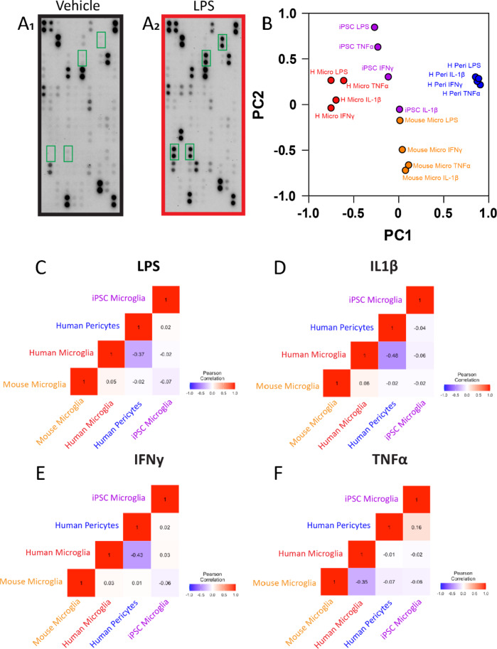
Microglia perform key homeostatic functions to protect the central nervous system (CNS). However, in many brain disorders their protective functions are abrogated, contributing to disease progression. Therefore, studies of microglial function are critical to developing treatments for brain disorders. Different in vitro microglia models have been established, including primary human and rodent cells, induced pluripotent stem cell (iPSC)-derived models, and immortalised cell lines. However, a direct comparative analysis of the phenotypic and functional characteristics of these models has not been undertaken. Accurate modelling of human microglia in vitro is critical for ensuring the translatability of results from the bench to the brain. Therefore, our study aimed to characterise and compare commonly utilised in vitro microglia models. We assessed four established microglia models: primary human microglia, human iPSC-derived microglia, the human microglial clone 3 (HMC3) cell line, and primary mouse microglia, with primary human brain pericytes acting as a negative control. Primary human microglia, iPSC-derived microglia, and mouse microglia stained positive for myeloid-cell markers (Iba1, CD45 and PU.1), while HMC3 cells only stained positive for mural-cell markers (PDGFRβ and NG2). Distinct secretomes were observed in all cell models in response to inflammatory treatment, with iPSC-derived microglia showing the most significant inflammatory secretions. Notably, nitric oxide was only secreted by mouse microglia. Although all cell types exhibited phagocytic capacity, primary human microglia and iPSC-derived microglia displayed significantly higher levels of phagocytosis. Overall, comparative analysis revealed notable differences between human microglia, iPSC-derived microglia, HMC3 cells and mouse microglia. Such differences should be considered when using these models to study human brain diseases. Experimental findings obtained from mouse models or cell lines should ultimately be cross validated to ensure the translatability of results to the human condition.

Keywords: In vitro models; Comparative analysis; Human microglial clone 3 (HMC3); Microglia; iPSC-derived microglia.


Conflict of interest statement

Declarations. Ethics approval and consent to participate: Studies undertaken using human tissue were approved by the Northern Regional Ethics Committee (New Zealand), with informed consent obtained from all donors. All methods were carried out in accordance with approved guidelines. Competing interests: The authors declare no competing interests.

Figures

Fig. 1
Fig. 1
Cell culture of in vitro microglia models. Schematic diagram depicting the steps followed to establish isolated human microglia, iPSC-derived microglia, HMC3 cells, human pericytes and mouse microglia cultures (A). Representative images of cell cultures under phase-contrast microscopy (B1–5). ssDMEM = serum-supplemented DMEM, GFDMEM = growth factor supplemented DMEM, see methods.
Fig. 2
Fig. 2
Expression of common microglia and mural-cell markers by each cell model. Isolated human microglia, iPSC-derived microglia, HMC3 cells, pericytes, or mouse microglia were plated and fixed for immunocytochemical staining of common microglia and mural markers. Representative immunofluorescent images of isolated human microglia, iPSC-derived microglia, HMC3 cells, and human pericyte stained with microglial-specific markers Iba1, CD45 and PU.1 (A) or mural-specific markers PDGFRβ, αSMA and NG2 (B). Representative images of isolated mouse microglia stained with PU.1 and Iba1 (C) or PDGFRβ and αSMA (D). Scale bars = 50 μm.
Fig. 3
Fig. 3
Comparison of the chemokine and cytokine secretion profile of each cell model. Cells were treated with inflammatory mediators (LPS, IL1β, IFNγ, TNFα) or vehicle (0.1% BSA in PBS) for 24 h, and conditioned media collected for measurements of analytes using the Proteome Profiler Cytokine Array Kit. Representative blots from vehicle and LPS treated iPSC-derived microglia (A1,2). For comparative analysis, each cell model’s treatment conditions were normalised back to vehicle. PCA plot displaying the clustering of each cell models treatment conditions (B). Pearson correlation matrices comparing the secretome of each cell type in response to LPS (C) IL1β (D), IFNγ (E) and TNFα (F) treatment. Scale bar denotes correlation coefficients, coloured according to value (blue, − 1.0, negatively correlated, red, 1.0, positively correlated).
Fig. 4
Fig. 4
Cytometric bead array quantification of cell models cytokine secretion in response to stimuli. Primary human microglia, iPSC-derived microglia, human pericytes, and mouse microglia were treated with inflammatory mediators (LPS, IL1β, IFNγ, TNFα) or vehicle (0.1% BSA in PBS) for 24 h and conditioned media collected for cytometric bead array or Griess assay analysis. Cytokine secretion data are represented as heat maps for each cell model, scale bar log2 concentration in pg/ml/10,000 cells (AE). Quantified secretion of cytokines and chemokines by each cell model (E1 - E10) n = 3–5, pg/ml/10,000 cells. Quantified nitric oxide (NO) secretion (µM) (F). n = 3 independent cases, data presented as mean ± SEM. One-way ANOVA with Dunnett’s multiple comparison test comparing vehicle with each treatment within each cell type * = p < 0.05, ** = p < 0.01, *** = p < 0.001, **** = p < 0.0001.
Fig. 5
Fig. 5
Comparison of phagocytic activity across microglial models. Human microglia, iPSC-derived microglia, human pericytes, or mouse microglia were plated in 48-well plates and incubated with 1 μm FluoSpheres™ Polystyrene Microspheres for 5 min, 1 h, 2 h, or 4 h. Cells were washed thoroughly to remove uninternalised beads, collected by trypsinisation and stained with 7-AAD to allow for gating on live cells. Representative fluorescent image confirming uptake of fluorescent beads by CD45 + human microglia (A). Scale bar = 50 μm. Active phagocytosis of beads was determined by a rightward shift in FL1 intensity via flow cytometry (B). Percentage of phagocytic cells at each time point for the four cell models (C). Mean fluorescent intensity (MFI) of gated phagocytic cells for each cell model at each time point (D). n = 3 independent repeats for each cell model, data presented as mean ± SEM. Two-way ANOVA with Dunnett’s multiple comparison test comparing the percentage of phagocytic cells and MFI at each time point to that of human microglia. * = p < 0.05, ** = p < 0.01, *** = p < 0.001, **** = p < 0.0001. Shared cell model key for C and D.
Fig. 6
Fig. 6
Schematic summary comparing the five in vitro cell models.

Similar articles

Cited by

References

    1. Prinz, M., Masuda, T., Wheeler, M. A. & Quintana, F. J. Microglia and central nervous System-Associated Macrophages-From origin to disease modulation. Annu. Rev. Immunol.39, 251–277 (2021). - PMC - PubMed
    1. Lapenna, A., De Palma, M. & Lewis, C. E. Perivascular macrophages in health and disease. Nat. Rev. Immunol.18 (11), 689–702 (2018). - PubMed
    1. Mildenberger, W., Stifter, S. A. & Greter, M. Diversity and function of brain-associated macrophages. Curr. Opin. Immunol.76, 102181 (2022). - PubMed
    1. Kierdorf, K., Masuda, T., Jordão, M. J. C. & Prinz, M. Macrophages at CNS interfaces: ontogeny and function in health and disease. Nat. Rev. Neurosci.20 (9), 547–562 (2019). - PubMed
    1. Timmerman, R., Burm, S. M. & Bajramovic, J. J. An overview of in vitro methods to study microglia. Front. Cell. Neurosci.12, 242 (2018). - PMC - PubMed

Publication types

LinkOut - more resources