. 2025 Jul;124(1):54-65.
doi: 10.1111/mmi.15367. Epub 2025 May 4.

Luminal Phospholipase D Attacks Bacterial Membranes in Dictyostelium discoideum Phagosomes

Affiliations

Luminal Phospholipase D Attacks Bacterial Membranes in Dictyostelium discoideum Phagosomes

Otmane Lamrabet et al. Mol Microbiol. 2025 Jul.

Abstract

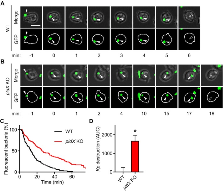
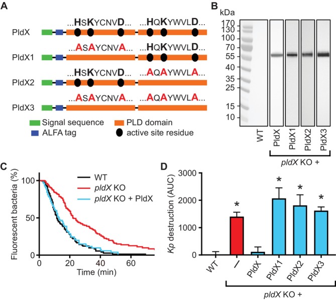
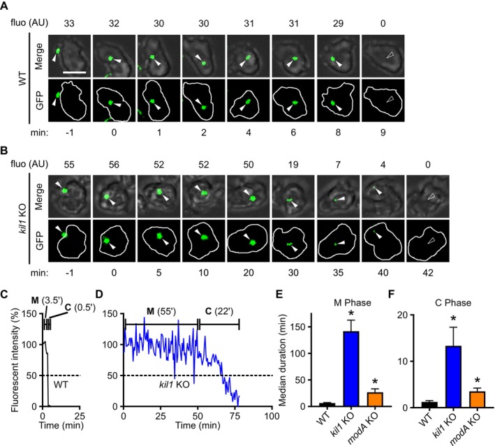
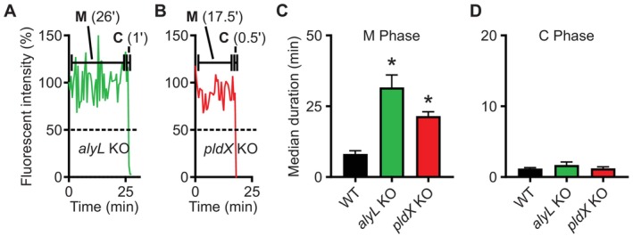
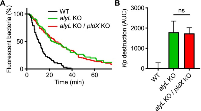
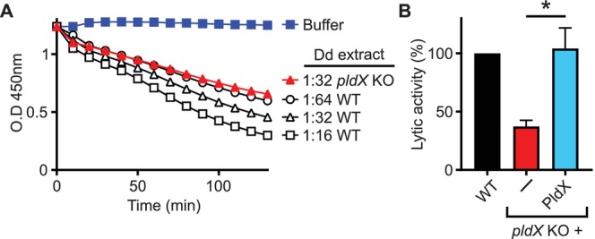
Phagocytic cells ingest bacteria and kill them in phagosomes. A variety of molecular mechanisms allow the killing and destruction of bacteria in phagosomes, but their complete list and relative importance remain poorly defined. Here we have used Dictyostelium discoideum amoebae as model phagocytic cells. Our results reveal that PldX, a luminal phospholipase D, plays an important role in the phagosomal destruction of ingested bacteria. Analysis of bacterial destruction in wild-type and pldX KO living cells suggests that PldX participates in the permeabilization of the bacterial membrane. The bacteriolytic activity of D. discoideum extracts was also measured in vitro: extracts from pldX KO cells exhibit significantly less bacteriolytic activity than wild-type cells, confirming the role of PldX in the lysis of bacterial membranes. These results identify luminal phospholipase D as a major player in the permeabilization of bacterial membranes in phagosomes.

Keywords: Dictyostelium discoideum; bacterial lysis; intracellular destruction; permeabilization of bacterial membrane; phospholipase D.


Conflict of interest statement

The authors declare no conflicts interest.

Figures

FIGURE 1
FIGURE 1
Three phospholipase D groups in Dictyostelium discoideum and in H. sapiens . Analysis of H. sapiens and D. discoideum sequence conservation as well as overall structure conservation revealed the existence of three groups of phospholipase D proteins: Mitochondrial, luminal, and secreted. A phylogenetic tree was obtained using maximum likelihood with the amino acid dataset. Numbers at the nodes represent the percentage of bootstrap support. A schematic representation of the three groups of PLDs is provided, with critical residues of the active sites marked in black.
FIGURE 2
FIGURE 2
Evolutionary conservation of cytosolic and luminal PLDs. Cytosolic (yellow squares) and luminal (red squares) PLD genes were identified and counted in a collection of eukaryotic species and in one archaeon. Several species of amoebozoa were also analyzed. Cytosolic and luminal PLDs appear to form two distinct groups. While cytosolic PLDs were found in all species analyzed, luminal PLDs were not found in the genomes of A. thaliana , S. cerevisiae , and E. histolytica .
FIGURE 3
FIGURE 3
PldX is required for efficient intracellular destruction of K. pneumoniae . To observe the internalization and the intracellular destruction of individual bacteria, D. discoideum cells were incubated with GFP‐expressing K. pneumoniae and observed for 2 h. (A) Successive images of a WT D. discoideum cell ingesting (t = 0) and destroying (t = 6 min) an individual K. pneumoniae bacterium. (B) Successive images of a pldX KO D. discoideum cell ingesting (t = 0) and destroying (t = 18 min) an individual K. pneumoniae bacterium. Scale bar 10 μm. (C) The time between ingestion and fluorescence extinction was determined for each bacterium and the probability of remaining fluorescent was represented as a function of time after ingestion in WT and pldX KO D. discoideum cells. The curves shown were obtained by pooling the results of 7 independent experiments. (D) Quantification of the normalized Area Under Curve (AUC) of independent experiments. In each independent experiment, a WT control was included, and its AUC subtracted from the AUC determined for the mutant cell (mean ± SEM. *: p < 0.05, Mann–Whitney test, WT: N = 7; n = 150, pldX KO: N = 7; n = 141).
FIGURE 4
FIGURE 4
Both active sites of PldX are necessary for its bacterial destruction activity. (A) PldX exhibits one signal sequence (green) and two PLD domains (orange). Each domain contains the catalytic HKD motif (HxKxxxxD). An ALFA tag (blue) was inserted at the N‐terminal end of the mature protein. The HKD motif was replaced with three alanines in the first PLD domain (PldX1), the second PLD domain (PldX2) or both (PldX3). (B) PldX, PldX1, PldX2 or PldX3 were expressed in pldX KO cells, and detected in cellular lysates by western blot using a recombinant antibody against the ALFA tag (full unedited gels are shown in Figure S4). WT cells were used as negative control. (C) Intracellular destruction of ingested K. pneumoniae was determined as described in Figure 3 in WT cells, pldX KO cells, and pldX KO cells expressing PldX. Expression of PldX in pldX KO cells restored rapid destruction of ingested K. pneumoniae . (D) Analysis of individual experiments confirmed that expression of PldX1, 2 and 3 did not restore efficient destruction of bacteria (mean ± SEM. *: p < 0.05, Mann–Whitney test; WT: N = 4, n = 107; pldX KO: N = 9, n = 232; PldX: N = 5, n = 109; PldX1: N = 6, n = 140; PldX2: N = 5, n = 132; PldX3: N = 6, n = 160).
FIGURE 5
FIGURE 5
The membrane (M) and cytosolic (C) phases of bacterial destruction are slower in kil1 KO and modA KO than in WT cells. WT (A) and kil1 KO (B) D. discoideum cells were incubated with GFP‐expressing K. pneumoniae and observed for 2 h. Scale bar 10 μm. For each ingested bacterium, the level of GFP fluorescence was determined as a function of time. In a typical WT cell (C), the fluorescence remained high for a few minutes (M phase 3.5 min) and then abruptly dropped (C phase 0.5 min). On the contrary, in a typical kil1 KO cell (D), the fluorescence remained high for a longer time (M phase 55 min) then gradually decreased (C phase, 22 min). The median duration of the M phase (E) and of the C phase (F) were determined in individual experiments (see Figure S5) for WT, kil1 KO, and modA KO cells (mean ± SEM. *: p < 0.05, Mann–Whitney test; WT: N = 14, n = 441 for M phase and n = 380 for C phase; kil1 KO: N = 5, n = 159 for M phase and n = 34 for C phase; modA KO: N = 11, n = 288 for M phase and n = 194 for C phase).
FIGURE 6
FIGURE 6
AlyL and PldX are required for rapid bacterial permeabilization. D. discoideum cells were incubated with GFP‐expressing K. pneumoniae . The fluorescence level of ingested bacteria was measured as a function of time as described in the legend of Figure 5. A representative example of bacterial destruction in an alyL KO cell (A) and in a pldX KO cell (B) is shown. The median duration of the M phase (C) and of the C phase (D) was determined in several independent experiments (mean ± SEM. *: p < 0.05, Mann–Whitney test; WT: N = 10, n = 254 for M phase and n = 209 for C phase; alyL KO: N = 5, n = 114 for M phase and n = 88 for C phase; pldX KO: N = 5, n = 129 for M phase and n = 90 for C phase).
FIGURE 7
FIGURE 7
Functional relationships between AlyL and PldX. The intracellular destruction of K. pneumoniae was assessed in WT, alyL KO mutant cells, and alyL/pldX double KO cells. (A) The kinetics of intracellular bacterial destruction were identical in alyL KO and alyL/pldX double KO cells. (B) Analysis of individual experiments (mean ± SEM. *: p < 0.05, Mann–Whitney test; WT: N = 3, n = 90; alyL KO: N = 3, n = 90; alyL KO/pldX KO: N = 6, n = 180).
FIGURE 8
FIGURE 8
Bacteriolytic activity is lower in extracts from pldX KO than from WT cells. (A) Bacteria were incubated at pH 2 in the presence of D. discoideum extracts, and the bacterial lysis was assessed by measuring light absorbance at 450 nm. Bacteriolytic activity was lower in diluted extracts from WT cells. Bacteriolytic activity was lower in extracts from pldX KO cells (red) than in extracts from WT cells. (B) Quantification of individual experiments revealed a significantly lower bacteriolytic activity in extracts from pldX KO cells. Expression of PldX restored a full lytic activity. (mean ± SEM. *: p < 0.05, Mann–Whitney test, N = 9 pldX KO; N = 3 for pldX KO + PldX).

Similar articles

References

    1. Alibaud, L. , Cosson P., and Benghezal M.. 2003. “ Dictyostelium discoideum Transformation by Oscillating Electric Field Electroporation.” BioTechniques 35: 78–83. - PubMed
    1. Ayadi, I. , Lamrabet O., Munoz‐Ruiz R., Jauslin T., Guilhen C., and Cosson P.. 2024. “Extracellular and Intracellular Destruction of Pseudomonas aeruginosa by Dictyostelium discoideum Phagocytes Mobilize Different Antibacterial Mechanisms.” Molecular Microbiology 121: 69–84. - PubMed
    1. Bodinier, R. , Leiba J., Sabra A., et al. 2020. “LrrkA, a Kinase With Leucine‐Rich Repeats, Links Folate Sensing With Kil2 Activity and Intracellular Killing.” Cellular Microbiology 22: e13129. - PMC - PubMed
    1. Bozzaro, S. 2013. “The Model Organism Dictyostelium discoideum .” In Methods in Molecular Biology, vol. 983, 17–37. Humana Press. - PubMed
    1. Choi, S. Y. , Huang P., Jenkins G. M., Chan D. C., Schiller J., and Frohman M. A.. 2006. “A Common Lipid Links Mfn‐Mediated Mitochondrial Fusion and SNARE‐Regulated Exocytosis.” Nature Cell Biology 8: 1255–1262. - PubMed

Substances