Review
. 2025 Jul;47(7):e70016.
doi: 10.1002/bies.70016. Epub 2025 May 4.

Mitochondria and Endoplasmic Reticulum Contact Site as a Regulator of Proteostatic Stress Responses in Neurodegenerative Diseases

Affiliations
Review

Mitochondria and Endoplasmic Reticulum Contact Site as a Regulator of Proteostatic Stress Responses in Neurodegenerative Diseases

Seiji Watanabe et al. Bioessays. 2025 Jul.

Abstract

Recent evidence indicates that the mitochondria-endoplasmic reticulum (ER) contact site is a novel microdomain essential for cellular homeostasis. Various proteins are accumulated at the mitochondria-associated membrane (MAM), an ER subcomponent closely associated with the mitochondria, contributing to Ca2+ transfer to the mitochondria, lipid synthesis, mitochondrial fission/fusion, and autophagy. These functions are disrupted in the diseases, particularly in neurodegenerative diseases such as amyotrophic lateral sclerosis (ALS) and Alzheimer's disease. In this review, we summarize the disruption of protein homeostasis in various neurodegenerative diseases, present recent works on the mechanisms of MAM aberration, including ours mainly focused on ALS, and then discuss challenges and prospects for future MAM-targeted therapies in neurodegenerative diseases.

Keywords: mitochondria‐associated membranes; neurodegenerative diseases; protein homeostasis.


Conflict of interest statement

The authors declare no conflicts of interest.

Figures

FIGURE 1
FIGURE 1
Proteostatic regulation at the MAM. Various proteins contributing cellular proteostasis, such as autophagy, ER‐associated protein degradation (ERAD), and ER stress response, are accumulated at the MAM. BiP, binding immunoglobulin protein/glucose regulated protein‐78; Der1, Derlin‐1; eIF2α, elongation initiation factor‐2α; ER, endoplasmic reticulum; MAM, mitochondria‐associated membrane; MFN2, mitofusin‐2; PERK, protein kinase R‐like endoplasmic reticulum kinase; RING, RING finger domain; STX17, syntaxin‐17; Ub, ubiquitin.
FIGURE 2
FIGURE 2
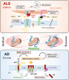
Schematic diagrams of the disease‐specific mitochondria‐associated membrane (MAM) alteration in amyotrophic lateral sclerosis (ALS) and Alzheimer's disease (AD). In ALS, the MAM is generally disrupted. Loss‐of‐function of the MAM‐specific ALS‐related protein, σ1R, and/or accumulation of misfolded mutant Cu/Zn superoxide dismutase (SOD1) compromises the interaction between inositol triphosphate receptor (IP3R) and voltage‐dependent anion channel 1 (VDAC1), resulting in decreased mitochondrial Ca2+ transfer and cytoplasmic Ca2+ leakage. Deregulated TAR DNA‐binding protein 43 (TDP‐43), a ubiquitously accumulated protein in ALS, causes activation of glycogen synthetase kinase 3β (GSK‐3β) to disrupt the interaction between vesicle‐associated membrane protein‐associated protein B (VAPB) and protein tyrosine phosphatase interacting protein 51 (PTPIP51) directly tethering the mitochondria and ER. On the other hand, in AD, accumulation of APP‐C99, a precursor of amyloid‐β (Aβ) processed by beta‐site amyloid precursor protein cleaving enzyme 1 (BACE1), enhances cholesterol and/or sphingolipids into the MAM. Increased cholesterol/sphingolipids contribute stabilization of the lipid microdomains including the MAM, resulting in excess MAM formation. The excess MAM further recruits BACE1 into the MAM to induce Aβ processing, which leads the formation of Aβ plaques.
FIGURE 3
FIGURE 3
The MAM is essential for TANK‐binding kinase 1 (TBK1)‐mediated stress response. Schematic illustration of TBK1‐mediated stress response dependent on the MAM. Our recent study [32] revealed that TBK1 is recruited into the MAM dependent on the stress‐dependent ubiquitination induced by autocrine motility factor receptor (AMFR/gp78). AMFR was anchored at the MAM via interaction with sigma 1 receptor (σ1R), an MAM‐specific chaperone protein. The recruited TBK1 was activated by its dimerization, leading to autophagic degradation of the translational components. This process promoted stress granule formation, contributing to improved stress tolerance in vitro and in vivo. CUE, coupling of ubiquitin to ER degradation domain; G3BP, GAP SH3‐binding protein 1; MAM, mitochondria‐associated membrane; RING, RING finger domain; Ub, ubiquitin; UBD, ubiquitin‐binding domain.

Similar articles

References

    1. Sassano M. L., Felipe‐Abrio B., and Agostinis P., “ER‐Mitochondria Contact Sites; a Multifaceted Factory for Ca2+ Signaling and Lipid Transport,” Frontiers in Cell and Developmental Biology 10 (2022): 988014. - PMC - PubMed
    1. Wilson E. L. and Metzakopian E., “ER‐Mitochondria Contact Sites in Neurodegeneration: Genetic Screening Approaches to Investigate Novel Disease Mechanisms,” Cell Death and Differentiation 28 (2021): 1804–1821. - PMC - PubMed
    1. Markovinovic A., Greig J., Martín‐Guerrero S. M., Salam S., and Paillusson S., “Endoplasmic Reticulum‐Mitochondria Signaling in Neurons and Neurodegenerative Diseases,” Journal of Cell Science 135 (2022): jcs248534. - PubMed
    1. Barazzuol L., Giamogante F., and Calì T., “Mitochondria Associated Membranes (MAMs): Architecture and Physiopathological Role,” Cell Calcium 94 (2021): 102343. - PubMed
    1. Vance J. E., “Phospholipid Synthesis in a Membrane Fraction Associated With Mitochondria,” Journal of Biological Chemistry 265 (1990): 7248–7256. - PubMed