

Integrated single-cell functional-proteomic profiling reveals a shift in myofibre specificity in human nemaline myopathy: A proof-of-principle study
- PMID: 40320980
- PMCID: PMC12126604
- DOI: 10.1113/JP288363
Integrated single-cell functional-proteomic profiling reveals a shift in myofibre specificity in human nemaline myopathy: A proof-of-principle study
Abstract
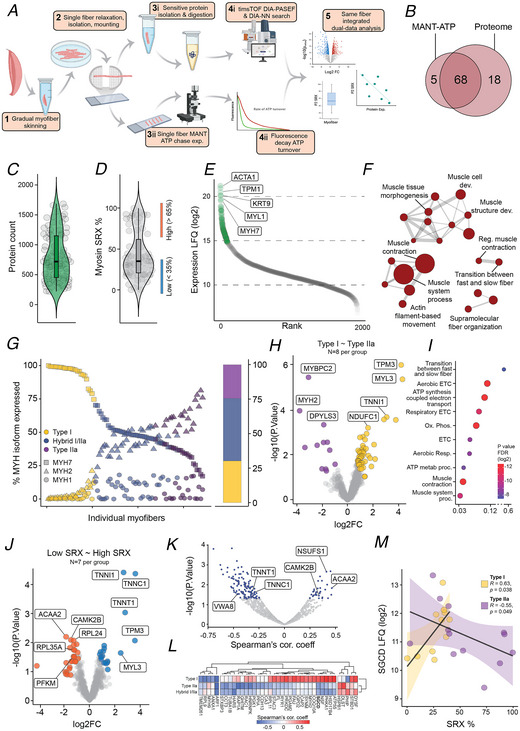
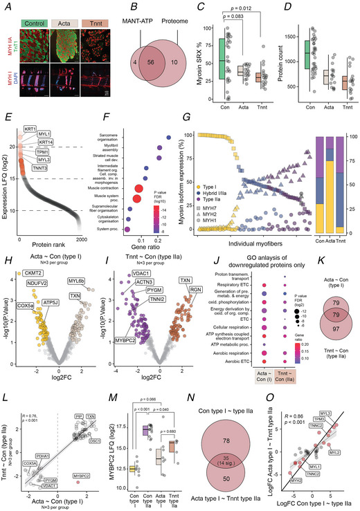
Skeletal muscle is a complex syncytial arrangement of an array of cell types and, in the case of muscle-specific cells (myofibres), subtypes. There exists extensive heterogeneity in skeletal muscle functional behaviour and molecular landscape at the cell composition, myofibre subtype and intra-myofibre subtype level. This heterogeneity highlights limitations in currently applied methodological approaches, which has stagnated our understanding of fundamental skeletal muscle biology in both healthy and myopathic contexts. Here we developed a novel approach that combines a fluorescence-based assay for the biophysical examination of the sarcomeric protein, myosin, coupled with same-myofibre high-sensitivity proteome profiling, termed single myofibre protein function-omics (SMPFO). Applying this approach as proof-of-principle we identify the integrated relationship between myofibre functionality and the underlying proteomic landscape that guides divergent, but physiologically important, behaviour in myofibre subtypes in healthy human skeletal muscle. By applying SMPFO to two forms of human nemaline myopathy (ACTA1 and TNNT1 mutations), we reveal significant reduction in the divergence of myofibre subtypes across both biophysical and proteomic behaviour. Collectively we demonstrate preliminary findings of SMPFO to support its use to study skeletal muscle with greater specificity, accuracy and resolution than currently applied methods, facilitating that advancement in understanding of skeletal muscle tissue in both healthy and diseased states. KEY POINTS: Skeletal muscle is a complex tissue made up of an array of cell and sub-cell types, with the resident muscle cell - myofibre - critical for contractile function. Although single myofibre studies have advanced, existing methods lack the precision for simultaneous multidata analysis, hindering developments in our understanding of skeletal muscle. We introduce single myofibre protein function-omics (SMPFO), a method enabling functional analysis of sarcomeric myosin alongside global protein abundance within the same myofibre. In healthy myofibres SMyoMFO reveals extensive biochemical diversity in myosin heads, correlating with the abundance of metabolic and sarcomeric proteins, including subtype-specific patterns in sarcoglycan delta (SGCD). In contrast SMyoMFO uniquely reveals a reduction in diversity of myosin function and the myofibre proteome in two forms of nemaline myopathy, highlighting disease-associated alterations. This innovative approach provides a robust framework for investigating myofibre regulation and dysfunction in skeletal muscle biology.
Keywords: human muscle; myopathy; myosin; proteomics.
© 2025 The Author(s). The Journal of Physiology published by John Wiley & Sons Ltd on behalf of The Physiological Society.
Conflict of interest statement
C.T.A.L. is an employee at Novo Nordisk A/S. Their contribution to this study was carried out prior to this employment and has no influence on the results presented or conclusions drawn in this study. This manuscript was originally submitted as a preprint to the bioRxiv preprint server for biology (Seaborne et al., 2024).
Figures
References
-
- Anderson, R. L. , Trivedi, D. V. , Sarkar, S. S. , Henze, M. , Ma, W. , Gong, H. , Rogers, C. S. , Gorham, J. M. , Wong, F. L. , Morck, M. M. , Seidman, J. G. , Ruppel, K. M. , Irving, T. C. , Cooke, R. , Green, E. M. , & Spudich, J. A. (2018). Deciphering the super relaxed state of human β‐cardiac myosin and the mode of action of mavacamten from myosin molecules to muscle fibers. Proceedings of the National Academy of Sciences of the United States of America, 115(35), E8143–e8152. - PMC - PubMed
-
- Angermueller, C. , Clark, S. J. , Lee, H. J. , Macaulay, I. C. , Teng, M. J. , Hu, T. X. , Krueger, F. , Smallwood, S. , Ponting, C. P. , Voet, T. , Kelsey, G. , Stegle, O. , & Reik, W. (2016). Parallel single‐cell sequencing links transcriptional and epigenetic heterogeneity. Nature Methods, 13(3), 229–232. - PMC - PubMed
-
- Argelaguet, R. , Clark, S. J. , Mohammed, H. , Stapel, L. C. , Krueger, C. , Kapourani, C. A. , Imaz‐Rosshandler, I. , Lohoff, T. , Xiang, Y. , Hanna, C. W. , Smallwood, S. , Ibarra‐Soria, X. , Buettner, F. , Sanguinetti, G. , Xie, W. , Krueger, F. , Göttgens, B. , Rugg‐Gunn, P. J. , Kelsey, G. , … Reik, W. (2019). Multi‐omics profiling of mouse gastrulation at single‐cell resolution. Nature, 576(7787), 487–491. - PMC - PubMed
-
- Bache, N. , Geyer, P. E. , Bekker‐Jensen, D. B. , Hoerning, O. , Falkenby, L. , Treit, P. V. , Doll, S. , Paron, I. , Müller, J. B. , Meier, F. , Olsen, J. V. , Vorm, O. , & Mann, M. (2018). A novel LC system embeds analytes in pre‐formed gradients for rapid, ultra‐robust proteomics. Molecular & Cellular Proteomics, 17(11), 2284–2296. - PMC - PubMed
MeSH terms
Substances
Grants and funding
- R347-2020-654/Lundbeck Foundation (Lundbeckfonden)
- R417-2022-1294/Lundbeck Foundation (Lundbeckfonden)
- R434-2023-311/Lundbeck Foundation (Lundbeckfonden)
- NNF21OC0070539/Novo Nordisk Fonden (NNF)
- NNF18CC0034900/Novo Nordisk Foundation Center for Basic Metabolic Research (NovoNordisk Foundation Center for Basic Metabolic Research)
LinkOut - more resources
Full Text Sources