

Dissecting Superficial Inferior Epigastric Veins Using a Tablet Navigation Device
- PMID: 40321336
- PMCID: PMC12047868
- DOI: 10.1097/GOX.0000000000006752
Dissecting Superficial Inferior Epigastric Veins Using a Tablet Navigation Device
Abstract
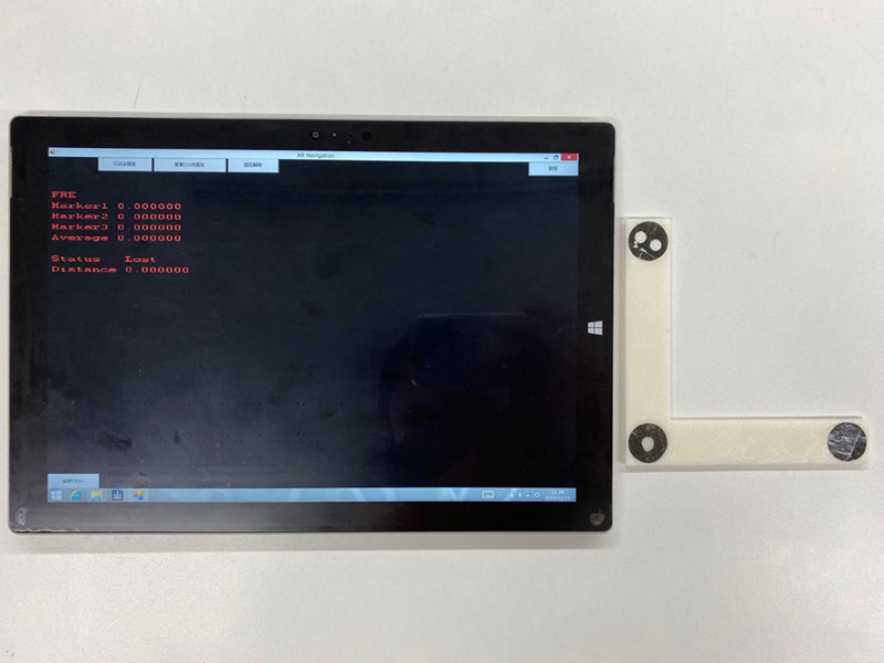
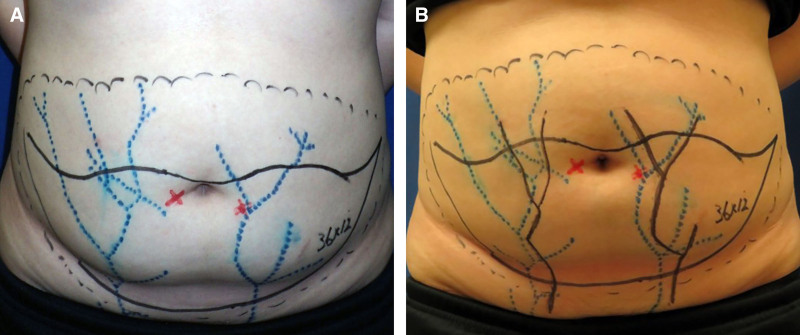
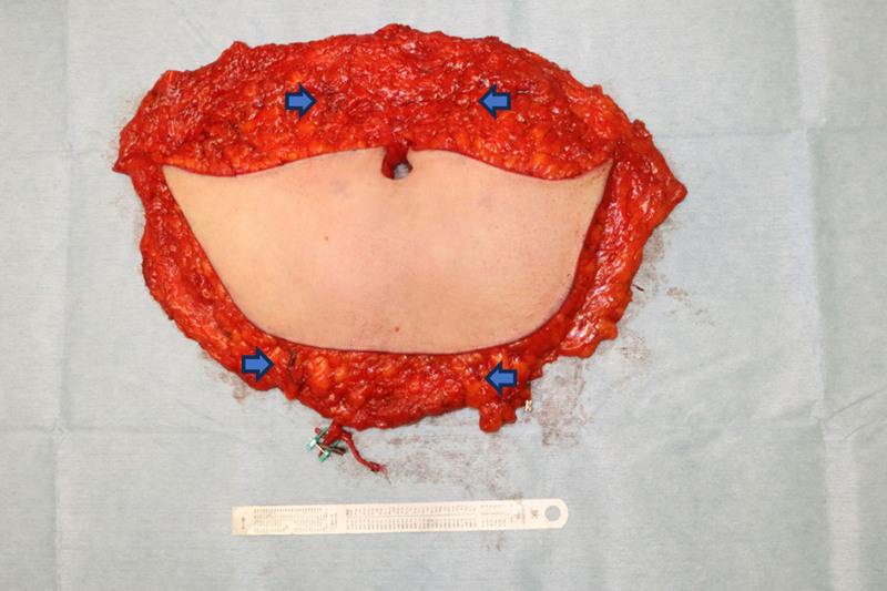
A preoperative understanding of the running pattern of blood vessels is important to safely raise flaps during surgery. The vascular anatomy from medical images indicates the entire vascular course and several perforator positions. We developed a navigation device using augmented reality technology to observe the running pattern of the vasculature collected from computed tomography angiography. Our navigation system was comprised of a tablet device (Surface Pro7), an L-shaped marker, and a custom-made software installed on the tablet device. The tablet device recognized three black and white circles, and generated vascular images of the patient's body that was filmed through the back camera of the device. Four deep inferior epigastric perforator (DIEP) artery flaps wherein the superficial inferior epigastric veins (SIEVs) were catheterized for flap monitoring, 2 DIEP flaps with superdrainage through the SIEV, and 1 superficial inferior epigastric artery flap were included in this study. Twelve SIEVs were safely dissected based on the SIEV markings of our navigation device. Despite an error of less than 1 cm, bilateral SIEVs were easily identified on the cranial and caudal sides using the image overlay system. Our device successfully navigated the superficial vascular dissection. In the future, we would like to apply this system to deep structure targets.
Copyright © 2025 The Authors. Published by Wolters Kluwer Health, Inc. on behalf of The American Society of Plastic Surgeons.
Conflict of interest statement
The authors have no financial interest to declare in relation to the content of this article. This study was supported by the JSPS KAKENHI (grant number: JP19K10037).
Figures
References
-
- Rozen WM, Ashton MW, Stella DL, et al. The accuracy of computed tomographic angiography for mapping the perforators of the DIEA: a cadaveric study. Plast Reconstr Surg. 2008;122:363–369. - PubMed
-
- Fukaya E, Kuwatsuru R, Iimura H, et al. Imaging of the superficial inferior epigastric vascular anatomy and preoperative planning for the SIEA flap using MDCTA. J Plast Reconstr Aesthet Surg. 2011;64:63–68. - PubMed
-
- Konoeda H, Uematsu M, Jumxiao N, et al. A trial to visualize perforators images from CTA with a tablet device: experience of operating on minipigs. Comput Assist Surg (Abingdon). 2022;27:120–127. - PubMed
-
- Lo S, Chapman P. The first worldwide use and evaluation of augmented reality (AR) in “patient information leaflets” in plastic surgery. J Plast Reconstr Aesthet Surg. 2020;73:1357–1404. - PubMed
-
- Vles MD, Terng NCO, Zijlstra K, et al. Virtual and augmented reality for preoperative planning in plastic surgical procedures: a systematic review. J Plast Reconstr Aesthet Surg. 2020;73:1951–1959. - PubMed
LinkOut - more resources
Full Text Sources