. 2025 Mar 24:90:e140-e150.
doi: 10.5114/pjr/200631. eCollection 2025.

Computed tomography radiomics combined with clinical parameters for hepatocellular carcinoma differentiation: a machine learning investigation

Affiliations

Computed tomography radiomics combined with clinical parameters for hepatocellular carcinoma differentiation: a machine learning investigation

Shijing Ma et al. Pol J Radiol. .

Abstract

Purpose: To evaluate the performance of a combined clinical-radiomics model using multiple machine learning approaches for predicting pathological differentiation in hepatocellular carcinoma (HCC).

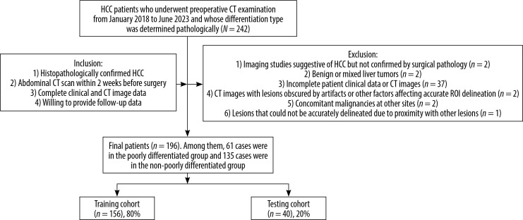
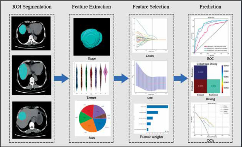
Material and methods: A total of 196 patients with pathologically confirmed HCC, who underwent preoperative computed tomography (CT) were retrospectively enrolled (training: n = 156; validation: n = 40). The modelling process included the folowing: (1) clinical model construction through logistic regression analysis of risk factors; (2) radiomics model development by comparing 6 machine learning classifiers; and (3) integration of optimal clinical and radiomic features into a combined model. Model performance was assessed using the area under the curve (AUC), calibration curves, and decision curve analysis (DCA). A nomogram was constructed for clinical implementation.

Results: Two clinical risk factors (BMI and CA153) were identified as independent predictors of differentiated HCC. The clinical model showed moderate performance (AUC: training = 0.705, validation = 0.658). The radiomics model demonstrated improved prediction capability (AUC: training = 0.840, validation = 0.716). The combined model achieved the best performance in differentiating HCC pathological grades (AUC: training = 0.878, validation = 0.747).

Conclusions: The integration of CT radiomics features with clinical parameters through machine learning provides a promising non-invasive approach for predicting HCC pathological differentiation. This combined model could serve as a valuable tool for preoperative treatment planning.

Keywords: computed tomography; hepatocellular carcinoma; machine learning; pathological grading; radiomics.


Figures

Figure 1
Figure 1
The workflow diagram showing the patient selection process. The study initially identified 242 HCC patients between January 2018 and June 2023. After applying inclusion and exclusion criteria, 196 patients were finally enrolled and divided into training (n = 156) and testing (n = 40) cohorts HCC – hepatocellular carcinoma, CT – computed tomography
Figure 2
Figure 2
Radiomics workflow showing the process of ROI segmentation, feature extraction, feature selection, and prediction model development Abbreviations: ROC – receiver operating characteristic, DCA – decision curve analysis
Figure 3
Figure 3
LASSO regression analysis results. A) Feature coefficient paths with optimal lambda value (λ = 0.0391). B) Mean squared error curve from 10-fold cross-validation. C) Coefficient values of the selected radiomic features. LASSO – least absolute shrinkage and selection operator, MSE – mean squared error, GLDM – grey level dependence matrix, GLCM – grey level co-occurrence matrix
Figure 4
Figure 4
Comparison of model performance in predicting HCC pathological differentiation. A) ROC curves comparing 6 machine learning algorithms in the training cohort, showing LightGBM achieved the highest AUC (0.840). B) ROC curves of the algorithms in the testing cohort, with LightGBM maintaining superior performance (AUC = 0.716). C) ROC curves comparing the clinical model (AUC = 0.705), radiomics model (AUC = 0.840), and combined model (AUC = 0.878) in the training cohort. D) Model comparison in the testing cohort, demonstrating maintained performance of the combined model (AUC = 0.747). E) Heat map of DeLong test results showing significant differences between the combined model and individual models (p < 0.05) ROC – receiver operating characteristic, AUC – area under the curve, CI – confidence interval, LightGBM – light gradient boosting machine, HCC – hepatocellular carcinoma
Figure 5
Figure 5
Performance evaluation and nomogram drawing of prediction models for pathological differentiation degree. A) Calibration curves comparing the agreement between predicted and observed probabilities in the training cohort for clinical, radiomics, and combined models. B) Decision curve analysis showing the net benefit of different models across various threshold probabilities. C) Nomogram integrating clinical and radiomics signatures for individualised prediction of hepatocellular carcinoma differentiation risk

Similar articles

References

    1. Mao XC, Shi S, Yan LJ, Wang HC, Ding ZN, Liu H, et al. . A model based on adipose and muscle-related indicators evaluated by CT images for predicting microvascular invasion in HCC patients. Biomark Res 2023; 11: 87. DOI: 10.1186/s40364-023-00527-z. - DOI - PMC - PubMed
    1. Jiang C, Cai YQ, Yang JJ, Ma CY, Chen JX, Huang L, et al. . Radiomics in the diagnosis and treatment of hepatocellular carcinoma. Hepatobiliary Pancreat Dis Int 2023; 22: 346-351. - PubMed
    1. Greten TF, Villanueva A, Korangy F, Ruf B, Yarchoan M, Ma L, et al. . Biomarkers for immunotherapy of hepatocellular carcinoma. Nat Rev Clin Oncol 2023; 20: 780-798. - PubMed
    1. Ozer Etik D, Suna N, Boyacioglu AS. Management of hepatocellular carcinoma: prevention, surveillance, diagnosis, and staging. Exp Clin Transplant 2017; 15: 31-35. - PubMed
    1. Martins-Filho SN, Paiva C, Azevedo RS, Alves VAF. Histological grading of hepatocellular carcinoma – a systematic review of literature. Front Med (Lausanne) 2017; 4: 193. DOI: 10.3389/fmed.2017.00193. - DOI - PMC - PubMed

LinkOut - more resources