[Preprint]. 2025 Apr 15:rs.3.rs-6248794.
doi: 10.21203/rs.3.rs-6248794/v1.

The transcription factor HHEX maintains glucocorticoid levels and protects adrenals from androgen-induced lipid depletion

Affiliations

The transcription factor HHEX maintains glucocorticoid levels and protects adrenals from androgen-induced lipid depletion

Typhanie Dumontet et al. Res Sq. .

Abstract

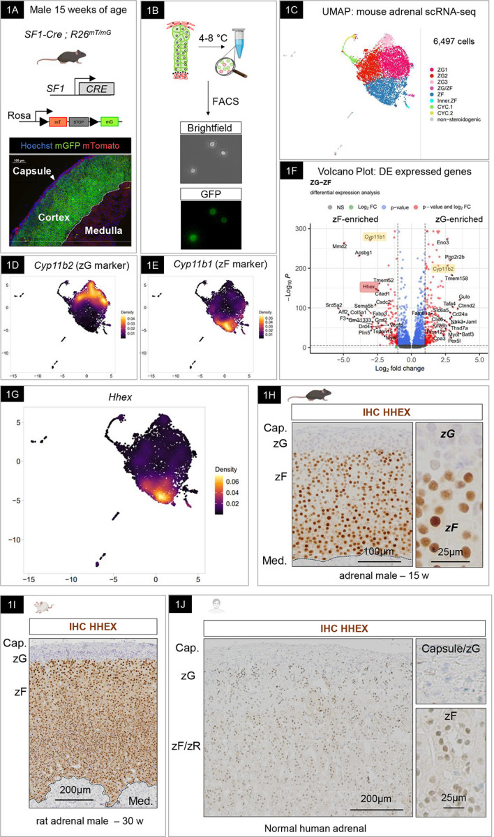
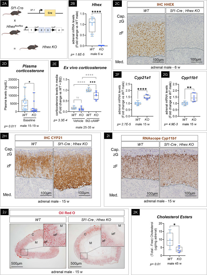
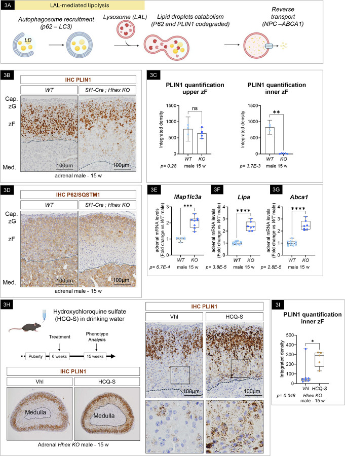
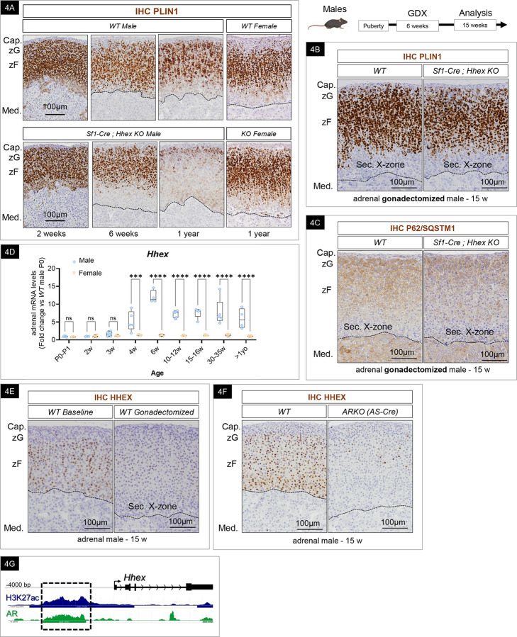
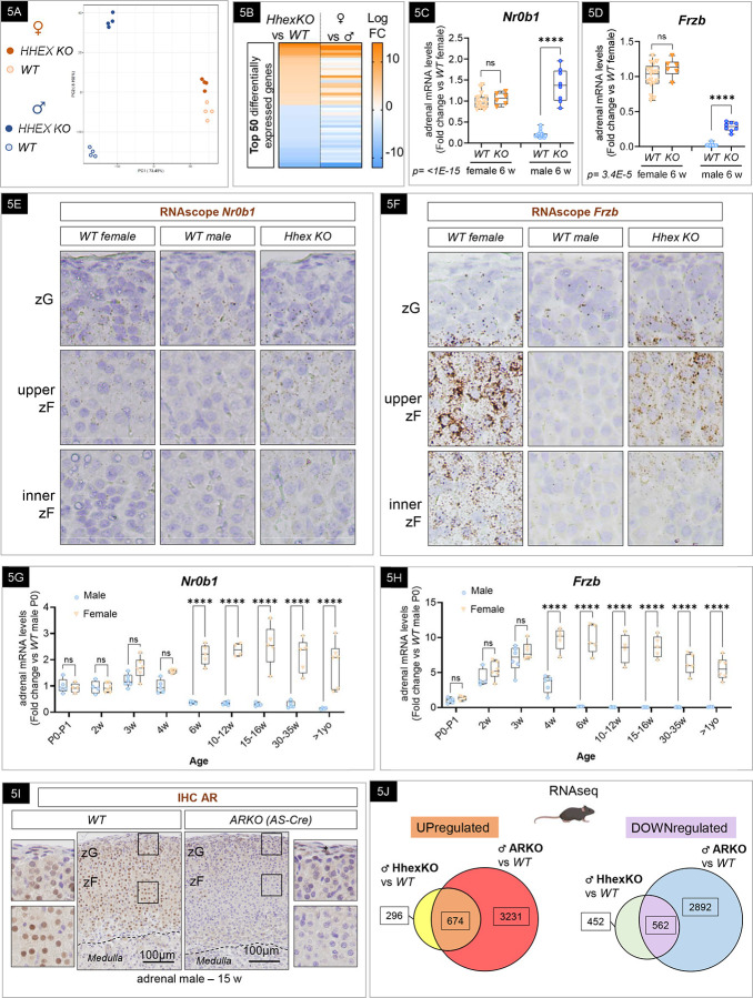
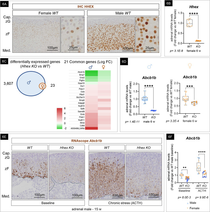
Glucocorticoid-producing cells of the adrenal cortex (i.e. zona fasciculata, zF) constitute the critical effectors of the hypothalamic-pituitary-adrenal axis, mediating the mammalian stress response. With glucocorticoids being essential for life, it is not surprising that zF dysfunction perturbs multiple organs that participate in optimizing cardiometabolic fitness. The zF forms a dynamic and heterogenous cell population endowed with the capacity to remodel through the engagement of both proliferative and differentiation programs that enable the adrenal to adapt and respond to diverse stressors. However, the mechanisms that sustain such differential responsiveness remain poorly understood. In this study, we resolved the transcriptome of the steroidogenic lineage by scRNA-seq using Sf1-Cre; Rosa mT/mG reporter mice. We identified HHEX, a homeodomain protein, as the most enriched transcription factor in glucocorticoid-producing cells. We developed new genetic mouse models to demonstrate that HHEX deletion causes glucocorticoid insufficiency in male animals. Molecularly, we demonstrated that HHEX is an androgen receptor (AR) target gene, shaping the sexual dimorphism of the adrenal gland by repressing the female transcriptional program at puberty, while also maintaining zF cholesterol ester content by protecting lipid droplets from androgen-induced-lipophagy. Moreover, our study revealed that, in both sexes, HHEX is crucial for maintaining the identity of the innermost adrenocortical cell subpopulation. Specifically, loss of HHEX impairs the expression of Abcb1b (P-glycoprotein/MDR1), an efflux pump regulating steroid export and cellular levels of xenobiotics. Together, these data demonstrate that HHEX serves as a multi-functional regulator of post-natal adrenal maturation that is potentiated by androgens.

Keywords: ACTH; HPA axis; PRH; Steroidogenic lineage; autophagy; cholesterol; corticosteroid; endocrine; lipid droplet; lipophagy; sexual dimorphism; stress response; testosterone; zona fasciculata.


Conflict of interest statement

Competing interest G.D.H. Founder and Board of Directors – Sling Therapeutics, Advisor – Orphagen Pharmaceuticals

Figures

Figure 1:
Figure 1:. scRNA-seq reveals heterogeneity within the steroidogenic lineage of the adrenal cortex and identified HHEX as a novel and highly conserved marker of GCs-producing cells.
(A) SF1-Cre mice were bred with RosamT/mG mice to label steroidogenic lineage with membrane Green Fluorescent Protein (mG). Non-recombined cells express Tomato (mT). Fluorescent image from a cryosection of an adult male SF1-Cre; mT/mG adrenal gland depicting endogenous mGFP (steroidogenic cells) and mTomato fluorescence. Hoechst was used to stain the nuclei. (B) Enzymatic and mechanic single-cell dissociation at low temperature, followed by FACS sorting. The image depicts GFP-positive cells after sorting. (C) UMAP representation of scRNA-seq dataset depicting steroidogenic cell populations in the adult male adrenal. zG: zona glomerulosa, zF: zona fasciculata, CYC: cycling. (D-E) UMAP representation of Cyp11b2 (D) and Cyp11b1 (E) expression in the scRNA-seq dataset. (F) Volcano Plot representing differentially expressed genes between zG and zF. Cyp11b1 and Cyp11b2 are highlighted in yellow and Hhex is highlighted in red. (G) UMAP representation of Hhex expression in the scRNA-seq dataset. (H-I-J) Expression of HHEX in the mouse (H), rat (I) and human (J) adrenal gland in brown by immunohistochemistry (IHC). Nuclei were stained in blue with hematoxylin.
Figure 2:
Figure 2:. Hhex KO male mice develop GC insufficiency.
(A) Schematic representation of the transgenic mouse models used to inactivate HHEX in adrenocortical cells using SF1-Cre-mediated recombination of exon 2 and 3 of Hhex gene. (B) Quantification of Hhex transcripts by RT-qPCR in 6-week-old WT (n=12) and SF1-Cre Hhex KO (n=8) male adrenals. Graph represents box plots with individual biological replicates. p-value (p) was calculated using a Mann-Whitney test. (C) Expression of HHEX in 6-week-old WT and SF1-Cre Hhex KO male adrenals in brown by immunohistochemistry (IHC). Nuclei were stained in blue with hematoxylin. Dotted lines represent the corticomedullary junction. WT: Wild Type, KO: Knockout, Cap.: Capsule, zG: zona glomerulosa, zF: zona fasciculata, Med.: medulla. (D) Corticosterone plasma levels of 15–19 week-old WT (n=21) and SF1-Cre; Hhex KO males (n=12) Graph represents box plots with individual biological replicates. p-value (p) was calculated using a Mann-Whitney test. (E) Ex vivo corticosterone production, released in culture media by adrenal explants after 2.5 hours incubation with 2.5mM of Bt2. WT (n=6) and SF1-Cre; Hhex KO males (n=7). Graph represents box plot with individual biological replicates. p-value (p) was calculated using a 2-way ANOVA followed by a Šídák’s multiple comparisons test. (F) Quantification of Cyp21a1 transcripts by RT-qPCR in 15-week-old WT (n=12) and SF1-Cre Hhex KO (n=7) male adrenals. Graph represents box plots with individual biological replicates. p-value (p) was calculated using a two-tailed unpaired t-test with Welch’s correction. (G) Quantification of Cyp11b1 transcripts by RT-qPCR in 15-week-old WT (n=12) and SF1-Cre Hhex KO (n=7) male adrenals. Graph represents box plots with individual biological replicates. p-value (p) was calculated using a two-tailed unpaired t-test with Welch’s correction. (H) CYP21A1 immunohistochemistry (IHC) in adrenals of WT and SF1-Cre; Hhex KO adrenals 15-week-old male mice. Nuclei were stained in blue with hematoxylin. Dotted lines represent the corticomedullary junction. WT: Wild Type, KO: Knockout, Cap.: Capsule, zG: zona glomerulosa, zF: zona fasciculata, Med.: medulla. (I) Cyp11b1 RNAscope in adrenals of WT and SF1-Cre; Hhex KO adrenals 15-week-old male mice. Nuclei were stained in blue with hematoxylin. Dotted lines represent the corticomedullary junction. WT: Wild Type, KO: Knockout, Cap.: Capsule, zG: zona glomerulosa, zF: zona fasciculata, Med.: medulla. (J) Oil Red O staining of 15-week-old WT and SF1-Cre; Hhex KO male adrenals. WT: Wild Type, KO: Knockout. Dotted lines represent corticomedullary junction. C: cortex, M.: medulla, zF, zona fasciculata, izF: inner zona fasciculata. (K) Cholesterol esters quantification in adrenals of 45-week-old WT and SF1-Cre; Hhex KO male. Graph represents box plots with individual biological replicates. p-value (p) was calculated using a two-tailed unpaired t-test with Welch’s correction.
Figure 3:
Figure 3:. Hhex KO adrenals display signs of activated lipophagy.
(A) Schematic of lipophagy also known as LAL-mediated lipolysis. (B) PLIN1 by immunohistochemistry (IHC) in adrenals of 15-week-old WT and SF1-Cre; Hhex KO adrenals. Nuclei were stained in blue with hematoxylin. Dotted lines represent the corticomedullary junction. WT: Wild Type, KO: Knockout, Cap.: Capsule, zG: zona glomerulosa, zF: zona fasciculata, Med.: medulla. (C) Quantification of PLIN1 immunohistochemistry in the upper zF and inner zF using imageJ. Graph represents mean with 95% Confidence Interval and individual biological replicates. p-value (p) was calculated using a two-tailed unpaired t-test with Welch’s correction. ns: non-significant. (D) P62/SQSTM1 immunohistochemistry (IHC) in adrenals of 15-week-old WT and SF1-Cre; Hhex KO adrenals. Nuclei were stained in blue with hematoxylin. Dotted lines represent the corticomedullary junction. WT: Wild Type, KO: Knockout, Cap.: Capsule, zG: zona glomerulosa, zF: zona fasciculata, Med.: medulla. (E-G) Quantification of Map1lc3a (E) (n=6 and 8), Lipa (F) (n=12 and 7), and Abca1 (G) (n=12 and 7) transcripts by RT-qPCR in 15-week-old WT and SF1-Cre; Hhex KO male adrenals. (H) PLIN1 immunohistochemistry (IHC) in adrenals of SF1-Cre; Hhex KO, Vehicle and hydroxychloroquine sulfate-treated 15-week-old male mice. Nuclei were stained in blue with hematoxylin. Dotted lines represent the corticomedullary junction. WT: Wild Type, KO: Knockout, Cap.: Capsule, zG: zona glomerulosa, zF: zona fasciculata, Med.: medulla. (I) Quantification of PLIN1 immunohistochemistry in the inner zF using imageJ. Vehicle n=7, HCQ-s n=5. Graph represents box plots with individual biological replicates. p-value (p) was calculated using a Mann-Whitney test.
Figure 4:
Figure 4:. Lipid depletion in Hhex KO male adrenals is androgens driven.
(A) PLIN1 immunohistochemistry (IHC) in adrenals of WT and SF1-Cre; Hhex KO adrenals at 2, 6, 15 weeks old and over 1-year-old male and female mice. Nuclei were stained in blue with hematoxylin. Dotted lines represent the corticomedullary junction. WT: Wild Type, KO: Knockout, Cap.: Capsule, zG: zona glomerulosa, zF: zona fasciculata, Med.: medulla. (B) PLIN1 immunohistochemistry (IHC) in adrenals of 15 week-old gonadectomized WT and SF1-Cre; Hhex KO male mice. Nuclei were stained in blue with hematoxylin. Dotted lines represent the corticomedullary junction. GDX: gonadectomy, WT: Wild Type, KO: Knockout, Cap.: Capsule, zG: zona glomerulosa, zF: zona fasciculata, Med.: medulla. Sec. X-zone: gonadectomy-induced secondary X-zone. (C) P62/SQSTM1 (lipophagy marker) immunohistochemistry (IHC) in adrenals of 15 week-old gonadectomized WT and SF1-Cre; Hhex KO male mice. Nuclei were stained in blue with hematoxylin. Dotted lines represent the corticomedullary junction. GDX: gonadectomy, WT: Wild Type, KO: Knockout, Cap.: Capsule, zG: zona glomerulosa, zF: zona fasciculata, Med.: medulla. Sec. X-zone: gonadectomy-induced secondary X-zone. (D) Timecourse of Hhex expression by RT-qPCR in male and female adrenal gland from birth to over 1 year old. Graph represents box plots with individual biological replicates. p-value (p) was calculated using a 2-way ANOVA followed by a Šídák’s multiple comparisons test. The number of biological replicates for each condition and a complete list of p-values are provided in the supplementary table related to Statistics (E) HHEX expression by immunohistochemistry (IHC) in the adrenals of 15-week-old WT intact and gonadectomized male adrenals. Nuclei were stained in blue with hematoxylin. Dotted lines represent the corticomedullary junction. GDX: gonadectomy, WT = Wild Type, Cap.: Capsule, zG: zona glomerulosa, zF: zona fasciculata, Med.: medulla. Sec. X-zone= gonadectomy-induced secondary X-zone. (F) HHEX expression by immunohistochemistry (IHC) in AS-Cre; ARKO. Nuclei were stained in blue with hematoxylin. Dotted lines represent the corticomedullary junction. WT: Wild Type, KO: Knockout, Cap.: Capsule, zG: zona glomerulosa, zF: zona fasciculata, Med.: medulla. Sec. X-zone: gonadectomy-induced secondary X-zone. (G) Histogram depicting the accumulation of androgen receptor (AR) CUT&Tag products obtained from adult male adrenal glands matching the Hhex promoter. H3K27ac is used to identify the open chromatin regions of the genome.
Figure 5:
Figure 5:. HHEX shapes the sexual dimorphism of the transcriptome of the zF.
(A) Principal Component Analysis (PCA) plots from bulk RNAseq analysis of 6-week-old WT and SF1-Cre; Hhex KO male and female adrenal glands (n=4 for each group). (B) Heatmaps of the top 50 differentially expressed genes between male SF1-Cre; Hhex KO and WT compared to dimorphic genes. (C-D) Nr0b1 (C) and Frzb (D) expression by RT-qPCR in the adrenal gland of 6-week-old female (▽) and male (), WT (n= 10 & 12) and SF1-Cre; Hhex KO (n= 6 & 8). Graph represents box plots with individual biological replicates. p-value (p) was calculated using a 2-way ANOVA followed by a Šídák’s multiple comparisons test. (E-F) Expression of Nr0b1 (E) and Frzb (E) by RNAscope in adrenal glands of 6-week-old WT female and WT and SF1-Cre; Hhex KO male. (G-H) Timecourse of Nr0b1 (G) and Frzb (H) expression by RT-qPCR in male () and female (▽) adrenal gland from birth to over 1 year old. Graph represents box plots with individual biological replicates. p-value (p) was calculated using a 2-way ANOVA followed by a Šídák’s multiple comparisons test. The number of biological replicates for each condition and a complete list of p-values are provided in the supplementary table related to Statistics (I) AR expression by immunohistochemistry (IHC) in 15-week-old WT and ARKO. Insets are focusing on capsule-zG cells (top) and zF cells (bottom). Nuclei are counterstained with hematoxylin. Dotted line represents the corticomedullary junction. Asterisk is pointing out AR-positive capsular cells in ARKO. zG: zona glomerulosa, zF = zona fasciculata. (J) Venn diagram of differentially expressed genes in SF1-Cre; Hhex KO and AS-Cre; ARKO compared to their respective WT.
Figure 6:
Figure 6:. HHEX is required for Abcb1b expression in the inner zF of both male and female adrenals.
(A) HHEX expression in adult female and male adrenal gland by immunohistochemistry (IHC). Nuclei are counterstained with hematoxylin. WT: Wild Type, Cap.: capsule, zG: zona glomerulosa, zF: zona fasciculata. (B) Quantification of Hhex transcripts by RT-qPCR in 6-week-old WT (n=10) and SF1-Cre; Hhex KO (n=6) female. Graphs represent individual biological replicates and the means with 95% confidence interval. p-value (p) is calculated using a two-tailed unpaired t-test with Welch’s correction. (C) Venn diagram representing the differentially expressed genes in SF1-Cre; Hhex KO compared to their respective WT (males in blue and females in orange) and heatmap of commonly differentially expressed genes in males and female SF1-Cre; Hhex KO. FC: Fold change. (D) Quantification of Abcb1b transcripts by RT-qPCR in 6-week-old WT (n=17 and 17) and SF1-Cre Hhex KO (n=8 and 6) male () and female (▽) adrenals. Graph represents box plots with individual biological replicates. p-value (p) is calculated using a two-tailed unpaired t-test with Welch’s correction. (E) Schematic of ACTH-induced chronic stress for 10 days in adult mice. Abcb1 expression in 15-week-old WT and SF1-Cre; Hhex KO male adrenals by RNAscope, at baseline and after chronic stress. Nuclei are stained in blue with hematoxylin. Dotted lines represent the corticomedullary junction. WT: Wild Type, KO: Knockout, Cap.: capsule, zG: zona glomerulosa, zF: zona fasciculata, Med: medulla (G) Quantification of Abcb1b transcripts by RT-qPCR in 15-week-old WT (n= 14 and 12) and SF1-Cre Hhex KO (n=14 and 8) male () and female (▽) adrenals. Graph represents box plots with individual biological replicates. p-value (p) is calculated using a 2-way ANOVA followed by a Šídák’s multiple comparisons test.

Similar articles

References

    1. Kuo T., McQueen A., Chen T.-C. & Wang J.-C. Regulation of Glucose Homeostasis by Glucocorticoids. Adv. Exp. Med. Biol. 872, 99–126 (2015). - PMC - PubMed
    1. Cain D. W. & Cidlowski J. A. Immune regulation by glucocorticoids. Nat. Rev. Immunol. 17, 233–247 (2017). - PMC - PubMed
    1. Dickmeis T. Glucocorticoids and the circadian clock. J. Endocrinol. 200, 3–22 (2009). - PubMed
    1. Marin M.-F. et al. Chronic stress, cognitive functioning and mental health. Neurobiol. Learn. Mem. 96, 583–595 (2011). - PubMed
    1. Shimba A. & Ikuta K. Glucocorticoids Regulate Circadian Rhythm of Innate and Adaptive Immunity. Front. Immunol. 11, 2143 (2020). - PMC - PubMed

Publication types

LinkOut - more resources