. 2025 Apr 30:20:5529-5549.
doi: 10.2147/IJN.S502724. eCollection 2025.

Novel Antigen-Presenting Cell-Targeted Nanoparticles Enhance Split Vaccine Immunity Through Microneedles Inoculation

Affiliations

Novel Antigen-Presenting Cell-Targeted Nanoparticles Enhance Split Vaccine Immunity Through Microneedles Inoculation

Xueliang Xiu et al. Int J Nanomedicine. .

Abstract

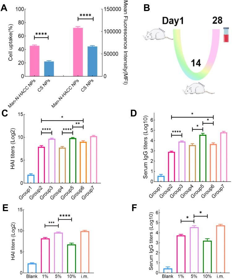
Aim/background: Despite their superior safety and widespread use, split vaccines typically suffer from reduced immunogenicity due to the lack of an intact viral structure. Targeting the mannose receptors on antigen-presenting cells (APCs) with nanoparticles (NPs) and delivering them via microneedles (MNs) offers a promising solution. We designed and synthesized NPs that could form complexes with split H1N1 antigens, and evaluated the immunogenicity after loading them into dissolvable microneedle arrays (dMAs).

Methods: Man-N-HACC was synthesized by conjugating mannose moieties to N-2-hydroxypropyl trimethyl ammonium chloride chitosan (N-HACC), followed by cross-linking with tripolyphosphate to form Man-N-HACC NPs. The NPs were characterized in terms of morphology, size, zeta potential, spatial orientation, macrophage internalization, and stability. The microstructure, mechanical strength, skin penetration capability, and release behavior of dMAs loaded with Man-N-HACC NPs/H1N1 complexes were investigated. Finally, the efficacy of dMAs was assessed in a rat model using ELISA and hemagglutination inhibition (HAI) assay.

Results: Characterization via Fourier transform infrared spectroscopy and nuclear magnetic resonance confirmed the synthesis of Man-N-HACC. The cross-linked generated Man-N-HACC NPs displayed uniform morphology and good stability over 28 days, along with confirmed spatial orientation of mannose ligands and macrophage internalization. The dMAs loaded with Man-N-HACC NPs/H1N1 exhibited mechanical robustness, capable of fully penetrating the skin and releasing nanovaccines. The increase in HAI titers and total IgG antibody levels in rat serum indicates the effectiveness of humoral immunity, and this effect only occurs after NPs formed post-crosslinking, rather than directly using raw nanomaterials, highlighting the critical role of the nanoparticle structure.

Conclusion: This study confirms that the delivery of Man-N-HACC NPs via dMAs provides a novel and promising approach for the administration of split influenza vaccines. Moreover, it underscores the great potential of nano-adjuvants in enhancing the efficacy of split vaccines.

Keywords: dissolvable microneedle arrays; mannose receptor; nanoparticles; split vaccine; target.


Conflict of interest statement

The authors declare that they have no known competing financial interests or personal relationships that could have appeared to influence the work reported in this paper.

Figures

None
Graphical abstract
Figure 1
Figure 1
Schematic representation of the immunization mechanism of dMAs patch loaded with Man-N-HACC NPs/H1N1. The NPs-split vaccine complex is released from the microneedle, and the mannose moiety on the surface of its NPs are specifically recognized and bound by the mannose receptor of APCs, generating endocytosis of the cell, which allows the vaccine particles to enter the cell and be processed and presented, resulting in a series of immune responses.
Figure 2
Figure 2
Synthesis and characterization of Man-N-HACC. (A) Synthesis route of N-HACC. GTMAC: glycidyl trimethylammonium chloride, N-HACC: N-2-hydroxypropyl trimethyl ammonium chloride chitosan. (B) Synthesis route of Man-N-HACC. R.T.: rotation. (C) FTIR spectra of CS, N-HACC, and Man-N-HACC. (D) 1H NMR spectra of N-HACC. (E) 1H NMR spectra of Man-N-HACC.
Figure 3
Figure 3
Physicochemical properties of the prepared Nps. (A) Impact of TPP concentration on the particle size and PDI of NPs. (B) Effect of TPP concentration on the Zeta potential of NPs. (C) TEM image of Man-N-HACC NPs, scale bar: 200 nm. (D) Particle size distribution of Man-N-HACC NPs. (E) Changes in particle size and PDI of Man-N-HACC NPs over 28 days at 4 °C. (F) Changes in Zeta potential of Man-N-HACC NPs over 28 days at 4 °C. (G) The size and PDI of H1N1, Man-N-HACC NPs and Man-N-HACC NPs/H1N1. (H) The Zeta potential of H1N1, Man-N-HACC NPs and Man-N-HACC NPs/H1N1. (I) Particle size distribution of Man-N-HACC NPs/H1N1.
Figure 4
Figure 4
Stability of Man-N-HACC NPs in MNs and characteristic changes upon binding with Con A (50 μg/mL). (A) Changes in particle size and PDI of Man-N-HACC NPs within MNs over 14 days at 4 °C. (B) Changes in Zeta potential of Man-N-HACC NPs within MNs over 14 days at 4 °C. (C) Schematic diagram of the lectin binding test. (D) Changes in particle size of Man-N-HACC NPs before and after binding with Con A (50 μg/mL). (E) Changes in particle size of CS NPs before and after binding with Con A (50 μg/mL). (F) Turbidity changes at 550 nm for Man-N-HACC NPs and CS NPs incubated with Con A (50 μg/mL).
Figure 5
Figure 5
SEM Imaging and mechanical characterization of NPs dMAs. SEM images of arrays (A), cross section (B) and tip local (C) without NPs-loaded dMAs. SEM images of 1% NPs-loaded dMAs array s tip local (D, scale bar: 50 μm) and amplified (E, scale bar: 5 μm). SEM images of 5% NPs-loaded dMAs tip local (F, scale bar: 50 μm) and amplified (G, scale bar: 5 μm). SEM images of 10% NPs-loaded dMAs tip local (H, scale bar: 50 μm) and amplified (I, scale bar: 5 μm). Insertion force of single microneedles loaded with 5% NPs (J) and mechanical properties of dMAs loaded with 1–10% NPs (K) tested by a high-sensitivity universal testing machine. Side (L) and top (M) views of NPs dMAs.
Figure 6
Figure 6
Performance of NPs dMAs penetrating simulated and real skins. (A) OCT image of 1% NPs dMAs penetrating the skin, scale bar: 600 μm. (B) OCT image of 5% NPs dMAs penetrating the skin, scale bar: 600 μm. (C) OCT image of 10% NPs dMAs s penetrating the skin, scale bar: 600 μm. (D) Results of the paraffin film puncture test for 5% NPs dMAs, scale bar: 1000 μm, 1–6 layer from top to bottom. (E) Illustration of the microneedle penetrating model skin with Rhodamine B as a model drug, scale bar: 600 μm. (F) Healing of skin pores within 12 hours, scale bar: 800 μm.
Figure 7
Figure 7
Immunization schedule and results of HAI and IgG antibody titer determination. (A) Quantitative analysis of cell internalization and intracellular fluorescence intensity for two different NPs. ****p < 0.0001. (B) Schematic representation of the immunization protocol. (C) HAI titers of different groups: group 1 (Blank dMAs containing PBS), group 2 (dMAs loaded with H1N1 antigen only), group 3 (H1N1/poly (I:C) dMAs), group 4 (Man-N-HACC/H1N1 dMAs), group 5 (5% Man-N-HACC NPs/H1N1 dMAs), group 6 (5% CS NPs/H1N1 dMAs), and group 7 (i.m. of H1N1). (D) IgG antibody titers of different groups. (E) HAI titers of microneedle patches with varying NPs concentrations: Blank (Blank dMAs containing PBS), 1% (1% Man-N-HACC NPs/H1N1 dMAs), 5% (5% Man-N-HACC NPs/H1N1 dMAs), and 10% (10% Man-N-HACC NPs/H1N1 dMAs). (F) IgG antibody titers of dMAs with varying NPs concentrations. *p < 0.05, **p < 0.01, ***p < 0.001, ****p < 0.0001.

Similar articles

References

    1. Uyeki TM, Hui DS, Zambon M, et al. Influenza. Lancet. 2022;400(10353):693–706. doi:10.1016/S0140-6736(22)00982-5 - DOI - PMC - PubMed
    1. Nir Y, Paz A, Sabo E, et al. Fear of injections in young adults: prevalence and associations. Am J Trop Med Hyg. 2003;68(3):341–344. doi:10.4269/ajtmh.2003.68.341 - DOI - PubMed
    1. Liu T, Luo G, Xing M. Biomedical applications of polymeric microneedles for transdermal therapeutic delivery and diagnosis: current status and future perspectives. Adv Ther. 2020;3(9). doi:10.1002/adtp.201900140 - DOI
    1. Zhang L, Xiu X, Li Z, et al. Coated porous microneedles for effective intradermal immunization with split influenza vaccine. ACS Biomater Sci Eng. 2023;9(12):6880–6890. doi:10.1021/acsbiomaterials.3c01212 - DOI - PubMed
    1. Song JM, Kim YC, E O, et al. DNA vaccination in the skin using microneedles improves protection against influenza. mol Ther. 2012;20(7):1472–1480. doi:10.1038/mt.2012.69 - DOI - PMC - PubMed

LinkOut - more resources