

Differential artery-vein analysis in OCTA for predicting the anti-VEGF treatment outcome of diabetic macular edema
- PMID: 40322014
- PMCID: PMC12047724
- DOI: 10.1364/BOE.557748
Differential artery-vein analysis in OCTA for predicting the anti-VEGF treatment outcome of diabetic macular edema
Abstract
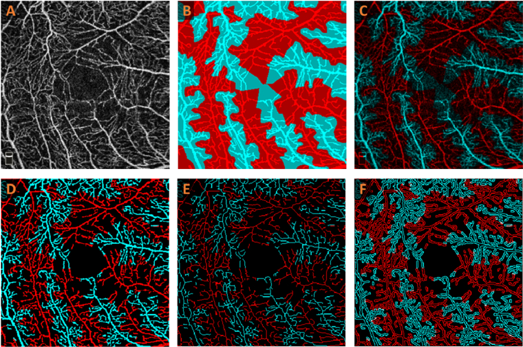
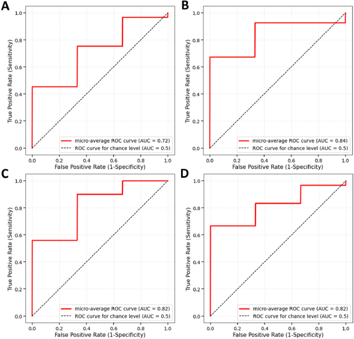
This study evaluates the role of differential artery-vein (AV) analysis in optical coherence tomography angiography (OCTA) for treatment outcome prediction of diabetic macular edema (DME). Deep learning AV segmentation in OCTA enabled the robust extraction of quantitative AV features, including perfusion intensity density (PID), blood vessel density (BVD), vessel skeleton density (VSD), vessel area flux (VAF), blood vessel caliber (BVC), blood vessel tortuosity (BVT), and vessel perimeter index (VPI). Support vector machine (SVM) classifiers were employed to predict changes in best-corrected visual acuity (BCVA) and central retinal thickness (CRT). Comparative analysis revealed that differential AV analysis significantly enhanced prediction performance, with BCVA accuracy improved from 70.45% to 86.36% and CRT accuracy enhanced from 68.18% to 79.55% compared to traditional OCTA analysis. These findings underscore the potential of AV analysis as a transformative tool for advancing personalized therapeutic strategies and improving clinical decision-making in managing DME.
© 2025 Optica Publishing Group.
Conflict of interest statement
No competing interest exists for any author.
Figures
Update of
- doi: 10.1364/opticaopen.28250855.
Similar articles
-
Differential artery-vein analysis improves the OCTA classification of diabetic retinopathy.Biomed Opt Express. 2024 May 22;15(6):3889-3899. doi: 10.1364/BOE.521657. eCollection 2024 Jun 1. Biomed Opt Express. 2024. PMID: 38867785 Free PMC article.
-
Application of optical coherence tomography angiography in the assessment of diabetic macular edema staging and laser photocoagulation efficacy.Photodiagnosis Photodyn Ther. 2024 Apr;46:104055. doi: 10.1016/j.pdpdt.2024.104055. Epub 2024 Mar 18. Photodiagnosis Photodyn Ther. 2024. PMID: 38508440
-
Differential Artery-Vein Analysis Improves the Performance of OCTA Staging of Sickle Cell Retinopathy.Transl Vis Sci Technol. 2019 Mar 26;8(2):3. doi: 10.1167/tvst.8.2.3. eCollection 2019 Mar. Transl Vis Sci Technol. 2019. PMID: 30941261 Free PMC article.
-
Quantitative Parameters Relevant for Diabetic Macular Edema Evaluation by Optical Coherence Tomography Angiography.Medicina (Kaunas). 2023 Jun 10;59(6):1120. doi: 10.3390/medicina59061120. Medicina (Kaunas). 2023. PMID: 37374324 Free PMC article. Review.
-
Optical Coherence Tomography Angiography of Macular Perfusion Changes after Anti-VEGF Therapy for Diabetic Macular Edema: A Systematic Review.J Diabetes Res. 2021 May 22;2021:6634637. doi: 10.1155/2021/6634637. eCollection 2021. J Diabetes Res. 2021. PMID: 34124270 Free PMC article.
References
LinkOut - more resources
Full Text Sources
Research Materials