. 2025;2(1):19.
doi: 10.1038/s44328-025-00038-x. Epub 2025 May 2.

A passive blood separation sensing platform for point-of-care devices

Affiliations

A passive blood separation sensing platform for point-of-care devices

Cameron Gilroy et al. NPJ Biosens. 2025.

Abstract

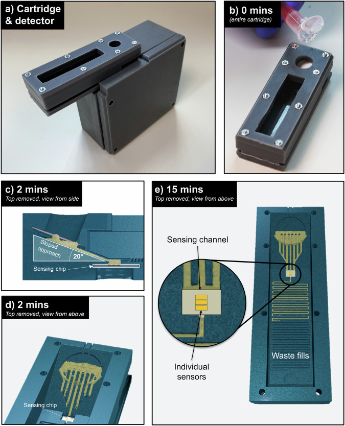
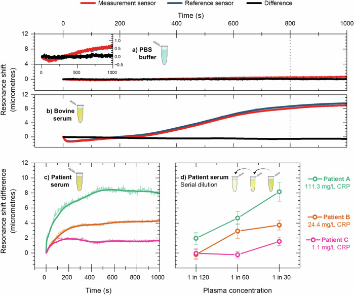
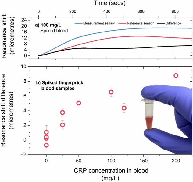
The blood test is one of the most performed investigations in clinical practice, with samples typically analysed in a centralised laboratory. Many of these tests monitor routine conditions that would benefit from a point-of-care approach, reducing the burden on practitioners, patients and healthcare systems. Such a decentralised model requires the development of sophisticated, yet easy-to-use technology; however, platforms that combine high-performance with low-cost and simplicity remain scarce. Moreover, most research papers only address a subset of requirements and study specific aspects in isolation. A systems approach that considers the interplay between each element of the technology is clearly required to develop a coherent solution. Here, we present such a systems approach in the context of testing for C-reactive protein (CRP), a commonly requested test in clinical practise that indicates inflammation and is particularly relevant for monitoring patients with chronic diseases, e.g. those with rheumatoid arthritis or who are undergoing cancer therapy. The approach we take here features an entirely passive microfluidic cartridge for blood separation, integrated with a high-performance sensing platform which we have tested in a real-world context. The device is compatible with a handheld detection unit and is simple to use yet can accurately detect CRP levels at clinically relevant levels.

Keywords: Applied optics; Biomedical engineering; Diagnostic markers; Lab-on-a-chip; Nanobiotechnology; Sensors and probes.


Conflict of interest statement

Competing interestsThe authors declare no competing interests.

Figures

Fig. 1
Fig. 1. Form and operation of the blood filtering device.
a 3D printed sensing platform and detection unit. b Blood sample is added to the inlet. c, d A sloped approach enables the gravity driven flow of blood plasma. e Plasma flows over the sensor and accumulates in a waste microfluidic channel.
Fig. 2
Fig. 2. Blood filtering capability of the device is measured by optical microscopy and absorption spectra measurements.
Optical micrographs of a Blood in buffer solution. Particle counting calculates 370 cells per 1000 µm2. b The same sample after passing through the cartridge showing 12 cells per 1000 µm2, a 97% reduction. c Plasma collected from a centrifuged sample showing 3 cells per 1000 µm. d Absorption spectra of blood and plasma solutions derived from different separation methods show that the cartridge approach performs almost as well as a centrifuge.
Fig. 3
Fig. 3. CRP concentration was measured in plasma solution derived from whole blood separated by the cartridge device and a centrifugation via enzyme linked immune immunosorbent assay (ELISA).
There is no significant loss of analyte
Fig. 4
Fig. 4. The sensing strategy of the device is functionalisation of a guided mode resonance sensor.
a Schematic of the silicon nitride ‘chirped’ guided mode resonance sensing chip. The sensor consists of two gratings in a ‘bowtie’ configuration to provide immunity to mechanical noise. The grating spacing here is exaggerated for illustrative purposes. b Experimental image captured of the sensor under monochromatic illumination. c Functionalisation of the top and bottom sensors with anti-CRP and goat IgG isotype antibodies, respectively, which we designate as measurement and reference sensors, respectively.
Fig. 5
Fig. 5. Sensing CRP in samples obtained from patient plasma samples.
a Resonance shift over time for the measurement and reference sensors in a PBS buffer solution. The error in the difference between sensors is approximately 0.10 microns. The inset shows same data on a smaller vertical scale. b In bovine serum, the measurement and reference sensors exhibit a binding curve associated with non-specific protein adsorption to the sensor surface however, the difference between sensors is also negligible, with an error of 0.70 microns. c The difference in resonance shift between the measurement and reference sensors for three patient serum samples in a 1 in 30 solution. d Serial dilution of patient serum samples showing increasing difference in resonance shift correlates with decreased dilution.
Fig. 6
Fig. 6. Sensing CRP in blood.
a Shift in the measurement and reference sensor resonance position and their difference for a 100 mg/L spiked blood sample. b Resonance shift difference for spiked fingerprick blood samples across a clinically relevant range.

Similar articles

References

    1. Future of POC and Rapid Testing. (Ipsos, Sermo).
    1. Lingervelder, D., Koffijberg, H., Kusters, R. & MJ, I. J. Health economic evidence of point-of-care testing: a systematic review. Pharmacoecon. Open5, 157–173 (2021). - PMC - PubMed
    1. Laurence, C. O. et al. Patient satisfaction with point-of-care testing in general practice. Br. J. Gen. Pr.60, e98–e104 (2010). - PMC - PubMed
    1. Kenaan, A. et al. Guided mode resonance sensor for the parallel detection of multiple protein biomarkers in human urine with high sensitivity. Biosens. Bioelectron.153, 112047 (2020). - PubMed
    1. Bakshi, S. et al. Nanophotonic and hydrogel-based diagnostic system for the monitoring of chronic wounds. Biosens. Bioelectron.242, 115743 (2023). - PubMed

LinkOut - more resources