Case Reports
. 2025 Apr 2;17(4):e81602.
doi: 10.7759/cureus.81602. eCollection 2025 Apr.

Multiple Myeloma With Leptomeningeal Involvement: A Study of Three Cases Exploring Diagnosis and Treatment Challenges

Affiliations
Case Reports

Multiple Myeloma With Leptomeningeal Involvement: A Study of Three Cases Exploring Diagnosis and Treatment Challenges

Jamil Abou Issa et al. Cureus. .

Abstract

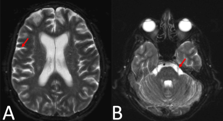
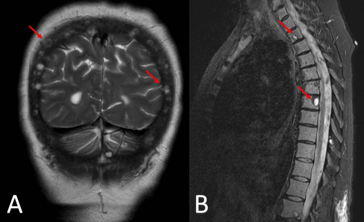
Central nervous system (CNS) involvement in multiple myeloma (MM) is a rare but serious complication that poses significant diagnostic and therapeutic challenges. This article presents three cases of leptomeningeal involvement in patients with MM, highlighting the diverse clinical presentations, diagnostic approaches, and treatment strategies employed. The first case is a 54-year-old female who, after initial treatment and autologous hematopoietic stem cell transplant, developed CNS disease. Similarly, a 73-year-old female developed leptomeningeal involvement with progressive neurological symptoms. The third case describes a 63-year-old female with immunoglobulin (Ig)A lambda MM who developed CNS disease after treatment with daratumumab and radiation. In all three cases, leptomeningeal enhancement and atypical plasma cells were identified in cerebrospinal fluid (CSF), with treatment strategies including intrathecal chemotherapy, systemic therapy, radiation, and stem cell transplantation. Despite aggressive management, including novel agents and supportive care, all patients had poor outcomes, with two transitioning to hospice care. The article reviews the limited literature on CNS-MM, noting the lack of standardized treatment protocols and the need for further research. As the survival of MM patients improves, the incidence of CNS involvement is expected to rise, making the development of targeted therapies essential. These cases underscore the urgent need for further investigation into novel treatment options and the importance of early diagnosis and comprehensive management of CNS-MM.

Keywords: leptomeningeal involvement; mm cns; multiple myeloma; multiple myeloma with cns involvement; multiple myeloma with extramedullary disease.


Conflict of interest statement

Human subjects: Consent for treatment and open access publication was obtained or waived by all participants in this study. Conflicts of interest: In compliance with the ICMJE uniform disclosure form, all authors declare the following: Payment/services info: All authors have declared that no financial support was received from any organization for the submitted work. Financial relationships: All authors have declared that they have no financial relationships at present or within the previous three years with any organizations that might have an interest in the submitted work. Other relationships: All authors have declared that there are no other relationships or activities that could appear to have influenced the submitted work.

Figures

Figure 1
Figure 1. Bone marrow morphology showing bone marrow effacement with plasma cells with plasmablastoid features
Panels A-C: Bone marrow showing effacement with plasma cells exhibiting plasmablastoid features at low, intermediate, and high magnifications, respectively.
Figure 2
Figure 2. MRI imaging of extra-axial mass in the right occipital lobe
Panel A: Axial T2-weighted MRI image showing a new extra-axial mass in the right occipital lobe, extending toward the cerebellum. Panel B: Sagittal T2-weighted MRI image of the same patient, highlighting the continuity of the extra-axial mass from the right occipital lobe to the cerebellum.
Figure 3
Figure 3. Coronal CT and PET imaging of multiple enhancing masses in the left abdomen
Panel A: Coronal CT scan of the abdomen and pelvis showing a conglomerate of multiple enhancing masses (red arrows) located in the left abdomen. Panel B: Coronal PET scan highlighting the same conglomerate of masses (red arrows) with increased metabolic activity, consistent with enhancing lesions. Both imaging modalities demonstrate the presence and localization of the masses in the left abdominal region.
Figure 4
Figure 4. Bone marrow infiltration by plasma cells with plasmablastic features
Panels A-C: Bone marrow showing effacement with plasma cells exhibiting plasmablastic features at low, intermediate, and high magnifications, respectively.
Figure 5
Figure 5. Axial brain MRI in T2-weighted FLAIR sequence demonstrating leptomeningeal enhancement and hyperintensity
Panel A: Axial T2-weighted fluid attenuated inversion recovery (FLAIR) MRI showing extensive abnormal hyperintense signal and leptomeningeal enhancement in the supratentorial compartment (red arrow), raising concern for leptomeningeal involvement. Panel B: Axial T2-weighted FLAIR MRI of the infratentorial compartment exhibiting similar extensive abnormal hyperintense signal and leptomeningeal enhancement (red arrow), further suggesting leptomeningeal disease.
Figure 6
Figure 6. PET scan imaging of hypermetabolic osseous lesions
Panel A: Positron emission tomography (PET) scan showing a lucent lesion in the right head of the humerus, indicated by the arrow, with a standardized uptake value (SUV) max of 4.0. Panel B: PET scan showing a lucent lesion in the right iliac bone (red arrow), depicting a lytic lesion with an SUV max of 5.1.
Figure 7
Figure 7. MRI imaging of osseous lesions in the calvarium and spine consistent with multiple myeloma
Panel A: T2-weighted coronal MRI scan of the brain displaying osseous lesions in the calvarium (red arrows). Panel B: T2/Short Tau Inversion Recovery (T2/STIR) sagittal MRI scan of the spine showing spinal lesions at the cervical and thoracic levels (red arrows).
Figure 8
Figure 8. CSF cytology showing atypical plasma cells
CSF: Ceebrospinal fluid.

References

    1. Expert review on soft-tissue plasmacytomas in multiple myeloma: definition, disease assessment and treatment considerations. Rosiñol L, Beksac M, Zamagni E, et al. Br J Haematol. 2021;194:496–507. - PubMed
    1. Extramedullary disease in multiple myeloma: a systematic literature review. Bladé J, Beksac M, Caers J, et al. Blood Cancer J. 2022;12:45. - PMC - PubMed
    1. Multiple myeloma with central nervous system relapse. Egan PA, Elder PT, Deighan WI, O'Connor SJ, Alexander HD. Haematologica. 2020;105:1780–1790. - PMC - PubMed
    1. Patterns of central nervous system involvement in relapsed and refractory multiple myeloma. Abdallah AO, Atrash S, Shahid Z, et al. Clin Lymphoma Myeloma Leuk. 2014;14:211–214. - PubMed
    1. Central nervous system involvement by multiple myeloma: a multi-institutional retrospective study of 172 patients in daily clinical practice. Jurczyszyn A, Grzasko N, Gozzetti A, et al. Am J Hematol. 2016;91:575–580. - PMC - PubMed

Publication types

LinkOut - more resources