

Detection, Prevention, and Treatment of Asymptomatic Myxoma in a Young Adult Patient: A Case Report
- PMID: 40322406
- PMCID: PMC12049076
- DOI: 10.7759/cureus.81649
Detection, Prevention, and Treatment of Asymptomatic Myxoma in a Young Adult Patient: A Case Report
Abstract
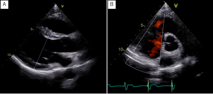
We present a case of myxoma originating from the right ventricular outflow tract. The patient was referred to our clinic after detecting a heart murmur at a routine school medical check-up, which is unique to Japan. He was treated with tumor resection and cryoablation to prevent recurrence. Cryoablation after tumor removal has the potential to prevent the recurrence of cardiac tumors. This case further emphasized the importance of regular school medical check-ups because it prevented sudden death.
Keywords: cardiac murmur; cardiac myxoma; cryoablation; school health check-up; ventricular outflow tract.
Copyright © 2025, Murai et al.
Conflict of interest statement
Human subjects: Consent for treatment and open access publication was obtained or waived by all participants in this study. Conflicts of interest: In compliance with the ICMJE uniform disclosure form, all authors declare the following: Payment/services info: All authors have declared that no financial support was received from any organization for the submitted work. Financial relationships: All authors have declared that they have no financial relationships at present or within the previous three years with any organizations that might have an interest in the submitted work. Other relationships: All authors have declared that there are no other relationships or activities that could appear to have influenced the submitted work.
Figures


Similar articles
-
[Myxoma originating from right ventricle found incidentally with cardiac murmur--a case report of surgical treatment].Nihon Kyobu Geka Gakkai Zasshi. 1993 Feb;41(2):288-94. Nihon Kyobu Geka Gakkai Zasshi. 1993. PMID: 8473798 Review. Japanese.
-
Left ventricular cardiac myxoma and sudden death in a dog.Acta Vet Scand. 2016 Jun 22;58(1):41. doi: 10.1186/s13028-016-0222-7. Acta Vet Scand. 2016. PMID: 27334273 Free PMC article.
-
Asymptomatic pediatric pulmonic valve myxoma involving the right ventricular outflow tract: a case report and review of the literature.J Heart Valve Dis. 2012 May;21(3):398-400. J Heart Valve Dis. 2012. PMID: 22808846
-
Right ventricular myxoma obstructing the outflow tract.Am Heart Hosp J. 2010 Winter;8(2):E118-21. doi: 10.15420/ahhj.2010.8.2.118. Am Heart Hosp J. 2010. PMID: 21928178
-
[Right ventricular myxoma--case report and review of the literature].Nihon Kyobu Geka Gakkai Zasshi. 1993 Jun;41(6):1069-73. Nihon Kyobu Geka Gakkai Zasshi. 1993. PMID: 8336036 Review. Japanese.
References
-
- Clinical presentation of left atrial cardiac myxoma. A series of 112 consecutive cases. Pinede L, Duhaut P, Loire R. Medicine (Baltimore) 2001;80:159–172. - PubMed
-
- Clinical characteristics and long term post-operative outcome of cardiac myxoma . Wu X, Yang D, Yang Z, Li J, Zhao Y, Wang K, Zhang R. https://pmc.ncbi.nlm.nih.gov/articles/PMC4943072/ EXCLI J. 2012;11:240–249. - PMC - PubMed
-
- Current challenges in the diagnosis and treatment of cardiac myxoma. Samanidis G, Khoury M, Balanika M, Perrea DN. Kardiol Pol. 2020;78:269–277. - PubMed
Publication types
LinkOut - more resources
Full Text Sources