

Temporal Artery Biopsies: Understanding the Low Positivity Rate
- PMID: 40322422
- PMCID: PMC12050073
- DOI: 10.7759/cureus.81714
Temporal Artery Biopsies: Understanding the Low Positivity Rate
Abstract
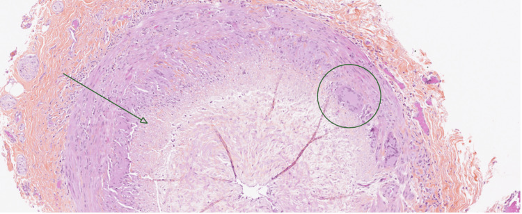
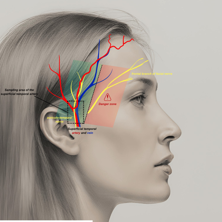
Objective We compared the yield of temporal artery biopsies with the clinical diagnoses made by referring physicians and determined the factors associated with a positive biopsy. Methods This is a monocentric, retrospective analytical study of patients treated between January 2021 and December 2024 who underwent temporal artery biopsy for suspected giant cell arteritis. The primary endpoint was the biopsy positivity rate. We also studied patient-related factors, symptoms, and clinical variables, including surgical and pathological factors, to identify those associated with a positive biopsy result. Results The study included 72 patients (40 females, 32 males). General health deterioration (OR = 3.71, p = 0.05) was significantly associated with a positive biopsy in the univariate analysis. The average length of the biopsy specimen after fixation was 13.4 mm. The positivity rate of temporal artery biopsies was 16.6% (n=12), while in 55.5% of cases (n=40), referring physicians ultimately diagnosed typical isolated Horton's disease (n=29, 40.2%) or isolated Horton's disease associated (n=10, 13.8%) with pseudo-polyarthritis rheumatica. Conclusion The anatomic-clinical discordance highlighted in our study supports findings from the literature. This can be explained by factors related to the pathology itself but also by a proactive diagnostic approach and variability in performing the surgical procedure. We have proposed avenues to refine the indications for this procedure and improve the performance of our technique. Temporal artery biopsy should be reserved for well-supported suspicions of Horton's disease. Sampling upstream from the bifurcation of the superficial temporal artery appears to offer the best reproducibility.
Keywords: general plastic surgery; good clinical practices; large-vessel vasculitis and gca; temporal arteritis; temporal artery biopsy.
Copyright © 2025, Girard et al.
Conflict of interest statement
Human subjects: Consent for treatment and open access publication was obtained or waived by all participants in this study. RGPD (European General Data Protection Regulation) issued approval MR004210220252. "In the research "Temporal Artery Biopsies : Understanding the Low Positivity Rate?", conducted by Mrs. Stella GIRARD and supervised by Professor Caroline FRANCOIS, a treatment record has been created and added to the GDPR register under registration number: MR004210220252. Please note that this document is available to the CNIL.". Animal subjects: All authors have confirmed that this study did not involve animal subjects or tissue. Conflicts of interest: In compliance with the ICMJE uniform disclosure form, all authors declare the following: Payment/services info: All authors have declared that no financial support was received from any organization for the submitted work. Financial relationships: All authors have declared that they have no financial relationships at present or within the previous three years with any organizations that might have an interest in the submitted work. Other relationships: All authors have declared that there are no other relationships or activities that could appear to have influenced the submitted work.
Figures
References
-
- 2012 revised International Chapel Hill Consensus Conference Nomenclature of Vasculitides. Jennette JC, Falk RJ, Bacon PA, et al. Arthritis Rheum. 2013;65:1–11. - PubMed
-
- Polymyalgia rheumatica and giant-cell arteritis. Salvarani C, Cantini F, Hunder GG. Lancet. 2008;372:234–245. - PubMed
-
- Operative technique: superficial temporal artery biopsy. Cahais J, Houdart R, Lupinacci RM, Valverde A. J Visc Surg. 2017;154:203–207. - PubMed
-
- The diagnosis and management of patients with giant cell arteritis. Gonzalez-Gay MA. https://pubmed.ncbi.nlm.nih.gov/15996050/ J Rheumatol. 2005;32:1186–1188. - PubMed
-
- Giant cell arteritis and polymyalgia rheumatica: an update. González-Gay MA, Pina T. http://link.springer.com/10.1007/s11926-014-0480-1. Curr Rheumatol Rep. 2015;17:6. - PubMed
LinkOut - more resources
Full Text Sources