

Necrotizing Fasciitis With Myonecrosis in a Diabetic Patient: Highlighting the Role of Early Detection and Management
- PMID: 40322434
- PMCID: PMC12050116
- DOI: 10.7759/cureus.81720
Necrotizing Fasciitis With Myonecrosis in a Diabetic Patient: Highlighting the Role of Early Detection and Management
Abstract
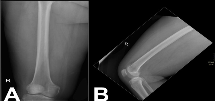
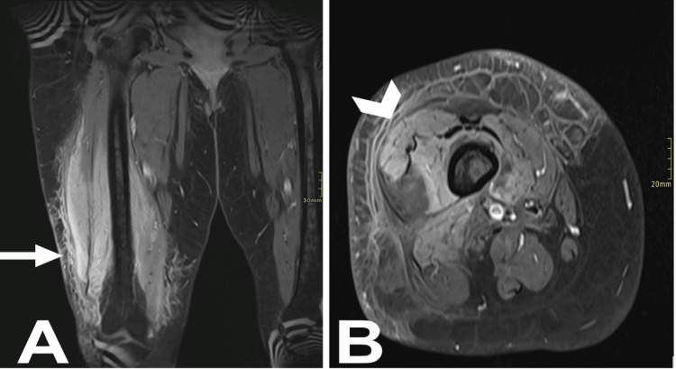
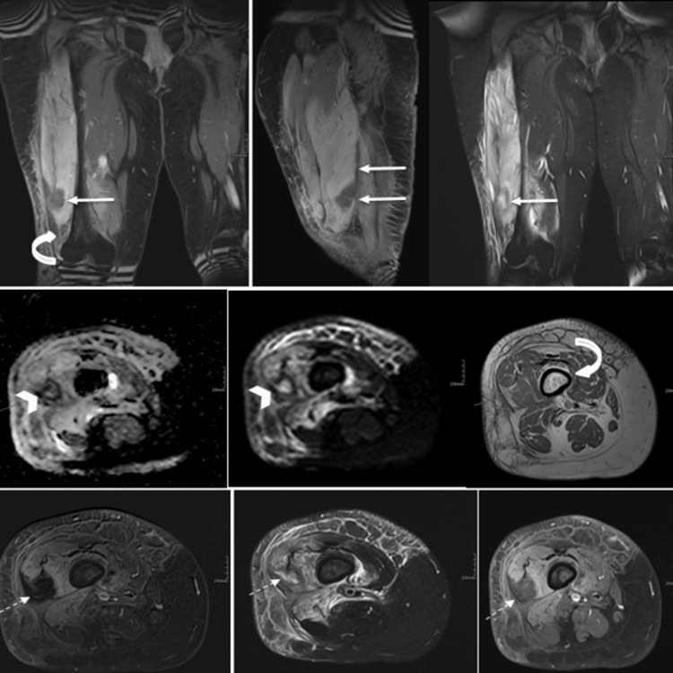
Necrotizing fasciitis (NF) is a life-threatening soft tissue infection that progresses rapidly and can lead to systemic complications. Myonecrosis, a severe complication of NF, involves muscle tissue death and often requires aggressive treatment. A 35-year-old female with diabetes mellitus, dyslipidemia, obesity, and a history of right breast cancer presented with acute, progressive right thigh pain, fever, and vomiting. Physical examination revealed local swelling, tenderness, warmth, and systemic signs of infection. Laboratory tests showed leukocytosis, elevated C-reactive protein, renal impairment, and hyponatremia. Contrast-enhanced MRI of the right thigh raised suspicion of NF with focal myonecrosis in the vastus lateralis and intermedius muscles. Surgical exploration and histopathology confirmed NF and myonecrosis. Debridement and broad-spectrum antibiotics, including vancomycin, meropenem, and clindamycin, were started. A second debridement and follow-up MRI showed improvement, with the patient recovering well and being discharged without complications. Early imaging, aggressive surgical intervention, and appropriate antibiotic therapy are critical in managing NF and myonecrosis, particularly in high-risk patients.
Keywords: diabetes; early detection; imaging; myonecrosis; necrotizing fasciitis.
Copyright © 2025, Alsaleh et al.
Conflict of interest statement
Human subjects: Consent for treatment and open access publication was obtained or waived by all participants in this study. Conflicts of interest: In compliance with the ICMJE uniform disclosure form, all authors declare the following: Payment/services info: All authors have declared that no financial support was received from any organization for the submitted work. Financial relationships: All authors have declared that they have no financial relationships at present or within the previous three years with any organizations that might have an interest in the submitted work. Other relationships: All authors have declared that there are no other relationships or activities that could appear to have influenced the submitted work.
Figures
Similar articles
-
Diabetic myonecrosis: likely an underrecognized entity.Orthopedics. 2014 Oct;37(10):e936-9. doi: 10.3928/01477447-20140924-91. Orthopedics. 2014. PMID: 25275984
-
A Rare and Unique Complication of Uncontrolled Type 2 Diabetes Mellitus: A Case Report and Literature Review of Spontaneous Diabetic Myonecrosis.Cureus. 2023 Apr 4;15(4):e37099. doi: 10.7759/cureus.37099. eCollection 2023 Apr. Cureus. 2023. PMID: 37168143 Free PMC article.
-
Case report: role of bedside ultrasonography in early diagnosis of myonecrosis rapidly developed in deep soft tissue infections.J Ultrasound. 2015 Feb 12;19(3):217-21. doi: 10.1007/s40477-015-0155-4. eCollection 2016 Sep. J Ultrasound. 2015. PMID: 27635157 Free PMC article.
-
Necrotizing fasciitis and gangrene of the upper extremity.Hand Clin. 1998 Nov;14(4):635-45, ix. Hand Clin. 1998. PMID: 9884900 Review.
-
[Necrotizing fasciitis in the head and neck region-three case reports and a review of the literature].HNO. 2020 Dec;68(12):935-943. doi: 10.1007/s00106-020-00899-w. HNO. 2020. PMID: 32617608 Review. German.
References
-
- LaChance A, Kroshinksy D. Fitzpatrick's Dermatology, 9th Edition. New York (NY): McGraw-Hill Education; 2019. Necrotizing fasciitis, necrotizing cellulitis, and myonecrosis.
-
- Necrotizing soft-tissue infections. Stevens DL, Bryant AE. N Engl J Med. 2017;377:2253–2265. - PubMed
Publication types
LinkOut - more resources
Full Text Sources
Research Materials