. 2025 Dec;17(1):2494717.
doi: 10.1080/19490976.2025.2494717. Epub 2025 May 5.

Defining enteric bacterial pathogenesis using organoids: Citrobacter rodentium uses EspC, an atypical mucinolytic protease, to penetrate mouse colonic mucus

Affiliations

Defining enteric bacterial pathogenesis using organoids: Citrobacter rodentium uses EspC, an atypical mucinolytic protease, to penetrate mouse colonic mucus

Yan Chen et al. Gut Microbes. 2025 Dec.

Abstract

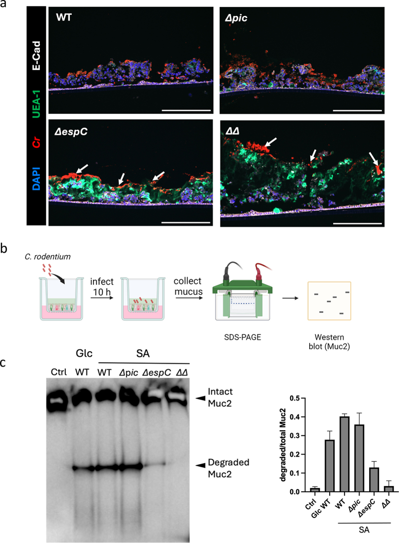
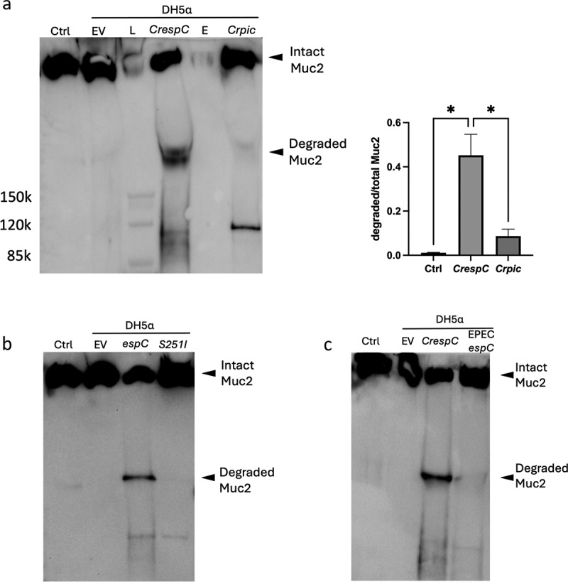
Enteric bacterial pathogens pose significant threats to human health; however, the mechanisms by which they infect the mammalian gut in the face of daunting host defenses remain to be fully defined. For the attaching and effacing (A/E) bacterial family member and murine pathogen Citrobacter rodentium, its virulence strategy appears to involve penetration of the colonic mucus barrier to reach the underlying epithelium. To better define these interactions, we grew colonoids under air-liquid interface (ALI) conditions, producing a thick mucus layer that mimicked in vivo mucus composition and glycosylation. C. rodentium's penetration of ALI-derived mucus was dramatically enhanced upon exposure to sialic acid, in concert with the secretion of two serine protease autotransporter of Enterobacteriaceae (SPATE) proteins, Pic and EspC. Despite Pic being a class II SPATE, and already recognized as a mucinase, it was EspC, a class I SPATE family member, that degraded ALI-derived mucus, despite class I SPATEs not previously shown to possess mucinase activity. Confirming this finding, E. coli DH5α carrying a plasmid that expresses C. rodentium-derived EspC was able to degrade the mucus. Moreover, recombinant EspC alone also displayed mucinolytic activity in a dose-dependent manner. Collectively, our results reveal the utility of ALI-derived mucus in modeling microbe-host interactions at the intestinal mucosal surface, as well as identify EspC as an atypical class I SPATE that shows significant mucinolytic activity toward ALI-derived mucus.

Keywords: Enteric pathogens; EspC; air-liquid interface; colonoids; mucus.


Conflict of interest statement

No potential conflict of interest was reported by the author(s).

Figures

Figure 1.
Figure 1.
The ALI model produces a physiologically relevant mucus layer. a, The experimental procedure to generate ALI-derived mucus, created with Biorender.com. b, The formation of the mucus layer after growing ALI cultures for 7 days, 14 days and 21 days. Arrows indicate the mucus layer. The average mucus thickness at 14 days and 21 days is 114 ± 28 µm, and 140 ± 25 µm, respectively. c, Immunostaining of Muc2-/- ALI (at 21 days). ALI cross-sections were stained with DAPI (blue), anti-Muc2 (green) and anti-E-cadherin (white). d, Proteomics analysis of gel-forming mucins present in mouse ALI-derived mucus. e-f, Pie chart of acidic (e) and various subsets (f) of mucin-type O-glycans in colonic ALI-derived mucus. g, MALII (red) staining of Muc2+/+ ALI culture. Scale bar, 200 µm.
Figure 2.
Figure 2.
The ALI model is able to recapitulate pathogen-mucus interaction in vivo. a, C. rodentium infection of Muc2+/+ ALI cultures for 6 h and 10 h. Scale bar, 200 µm. b, C. rodentium infection of Muc2-/- ALI culture for 6 h. ALI cross-sections were stained with anti-C. rodentium LPS (red), Ulex europaeus agglutinin-1 (UEA-1, green), DAPI (blue) and E-cadherin (white). Scale bar, 200 µm. White arrows indicate C. rodentium present in the mucus. Yellow arrows indicate C. rodentium close to IECs. Yellow arrowheads indicate cell sloughing.
Figure 3.
Figure 3.
Sialic acid enhances C. rodentium’s ability to degrade ALI-derived mucus and infect IECs. a, Diagram of mucus degradation assay in vitro, created with Biorender.com. b, Mucus degradation assay using the supernatant from C. rodentium grown in media containing 0.45% glucose (Glc) or sialic acid (SA). Mucus incubated with C. rodentium supernatants was loaded onto 3–8% Tris-acetate gels and run through electrophoresis. Proteins were visualized by western blot using an anti-Muc2 antibody. c, Quantification analysis of degraded Muc2 band to total Muc2 band, ***, p < 0.001; ****, p < 0.0001. d, Immunostaining of ALI cultures infected with C. rodentium in the presence of glucose or sialic acid. ALI cross-sections were stained with anti-C. rodentium LPS (red), UEA-1(green), DAPI (blue) and E-cadherin (white). Yellow arrows indicate C. rodentium close to IECs. Yellow arrowheads indicate cell sloughing. Scale bar, 200 µm.
Figure 4.
Figure 4.
A comparison of the ability of WT, Δpic, ΔespC and ΔpicΔespC C. rodentium strains to infect mouse ALI cultures and to degrade the ALI-derived mucus. a, The ALI cultures were infected with different C. rodentium strains, including WT, Δpic, ΔespC and ΔpicΔespC double mutant (ΔΔ). ALI cross-sections were stained with anti-C. rodentium LPS (red), UEA-1(green), DAPI (blue) and E-cadherin (white). White arrows indicate C. rodentium on the top of the mucus. Scale bar, 200 µm. b, Diagram of mucus degradation assay after C. rodentium infection, created with Biorender.com. ALI cultures were infected with different bacterial strains, followed by the collection of mucus from infected cultures. c, mucus from infected ALI was collected and run on a 3%-8% Tris-acetate gel, followed by western blot using an anti-Muc2 antibody.
Figure 5.
Figure 5.
Mucus degradation assay using protein secreted by SPATEs expressing E. coli DH5α. a, The degradation of mouse ALI-derived mucus by supernatant from E. coli DH5α expressing CrespC or Crpic. EV, empty vector; L, protein ladder; E, empty lane. Note, the anti-Muc2 antibody shows cross-activity against Pic (~110 kDa). *, p < 0.05. b, The degradation of mouse ALI-derived mucus by supernatant from E. coli DH5α expressing CrespC or CrespC-S251I. c, The degradation of mouse ALI-derived mucus by supernatant from E. coli DH5α expressing CrespC or EPECespC. Samples from the mucus degradation assay were reduced and run on a 3%-8% Tris-acetate gel, followed by western blot using an anti-C-terminus Muc2 antibody.
Figure 6.
Figure 6.
Recombinant CrEspC is able to degrade mouse ALI-derived mucus. a, The sequence information used to express recombinant CrEspC by E. coli BL21 Star (DE3) system, created with Biorender.com. SS, signal sequence. b, The degradation of mouse ALI-derived mucus by 0.05 µg purified CrEspC for 2 h, 8 h and 24 h. c, The degradation of mouse ALI-derived mucus by different amounts (0.0025, 0.0125, 0.05 µg) of purified CrEspC for 2 h. Samples were reduced and run on a 3%-8% Tris-acetate gel, followed by western blot using an anti-C-terminus Muc2 antibody.

References

    1. Croxen MA, Law RJ, Scholz R, Keeney KM, Wlodarska M, Finlay BB.. Recent advances in understanding enteric pathogenic Escherichia coli. Clin Microbiol Rev. 2013;26(4):822–19. doi: 10.1128/CMR.00022-13. - DOI - PMC - PubMed
    1. Lu L, Walker WA. Pathologic and physiologic interactions of bacteria with the gastrointestinal epithelium. Am J Clin Nutr. 2001;73(6):1124S–1130S. doi: 10.1093/ajcn/73.6.1124S. - DOI - PubMed
    1. Mundy R, Girard F, Fitzgerald AJ, Frankel G. Comparison of colonization dynamics and pathology of mice infected with enteropathogenic Escherichia coli, enterohaemorrhagic E. coli and citrobacter rodentium. FEMS Microbiol Lett. 2006;265(1):126–132. doi: 10.1111/j.1574-6968.2006.00481.x. - DOI - PubMed
    1. Silberger DJ, Zindl CL, Weaver CT. Citrobacter rodentium: a model enteropathogen for understanding the interplay of innate and adaptive components of type 3 immunity. Mucosal Immunol. 2017;10(5):1108–1117. doi: 10.1038/mi.2017.47. - DOI - PMC - PubMed
    1. Deng W, Puente JL, Gruenheid S, Li Y, Vallance BA, Vázquez A, Barba J, Ibarra JA, O’Donnell P, Metalnikov P, et al. Dissecting virulence: systematic and functional analyses of a pathogenicity island. Proc Natl Acad Sci, India, Sect B Biol Sci. 2004;101(10):3597–3602. doi: 10.1073/pnas.0400326101. - DOI - PMC - PubMed

Substances

LinkOut - more resources