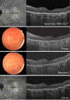
Imaging of Geographic Atrophy: A Practical Approach
- PMID: 40323558
- PMCID: PMC12167423
- DOI: 10.1007/s40123-025-01158-3
Imaging of Geographic Atrophy: A Practical Approach
Abstract
Geographic atrophy (GA) secondary to age-related macular degeneration is a chronic degenerative disease involving the retinal pigment epithelium, photoreceptors, and choriocapillaris leading to irreversible loss of visual function. Identification of imaging markers associated with GA development and progression has progressed over the past decades, moving from two-dimensional to three-dimensional imaging, as well as image interpretation using artificial intelligence. However, there is an open discussion about the "must-haves" for GA detection and follow-up as well as complementary imaging. This practical approach provides an overview of the advantages of key imaging modalities for GA and their applicability in clinical and experimental settings.
Keywords: Age-related macular degeneration; Fundus autofluorescence; Geographic atrophy; Imaging; Optical coherence tomography.
© 2025. The Author(s).
Conflict of interest statement
Declarations. Conflict of interest: Gregor S. Reiter: consultant for Apellis, Bayer, Boehringer Ingelheim, Espansione, Nordic Pharma, and Roche and received research support from RetInSight. Enrico Borrelli: consultant for Abbvie, Bayer, Hofmann La Roche, and Zeiss. Rosa Dolz-Marco: consultant for Heidelberg Engineering and received research support from Roche, Apellis, and IvericBio. Raymond Iezzi: consultant for Johnson & Johnson. Sophie J. Bakri: consultant for Abbvie, Adverum, Allergan, Amgen, Annexon, Apellis, Aviceda, Cholgene, Eyepoint, ilumen, Iveric bio, Kala, Genentech, La Science Neurotech, Novartis, Ocular Therapeutix, Opthea, Outlook, Pixium, Regenxbio, Regeneron, Rejuvitas, Revana, Roche, VoxelCloud, and Zeiss and research support from Lowy Medical Foundation and Regenxbio. Ethical approval: This article is based on previously conducted studies and does not contain any new studies with human participants or animals performed by any of the authors.
Figures


References
-
- Zarbin MA, Casaroli-Marano RP, Rosenfeld PJ. Age-related macular degeneration: Clinical findings, histopathology and imaging techniques. Cell-Based Therapy for Retinal Degenerative Disease. S. Karger AG 2014:1–32. - PubMed
LinkOut - more resources
Full Text Sources
Miscellaneous

