

The process of residual calcification following antiparasitic treatment in the pig model of neurocysticercosis is dynamic
- PMID: 40323912
- PMCID: PMC12052124
- DOI: 10.1371/journal.pntd.0013022
The process of residual calcification following antiparasitic treatment in the pig model of neurocysticercosis is dynamic
Abstract
Background: Calcified neurocysticercosis (NCC), the end stage of brain cysts of the pork tapeworm Taenia solium is a common cause of epilepsy. Calcified NCC lesions are not inert and represent potential epileptogenic foci. Understanding the mechanisms of residual calcification in NCC is hindered by the difficulty of accessing human brain biopsies. Since cyst degeneration can be induced by antiparasitic treatment (APT) in NCC-infected pigs, this study assessed the residual calcification process in this model at three time points after APT.
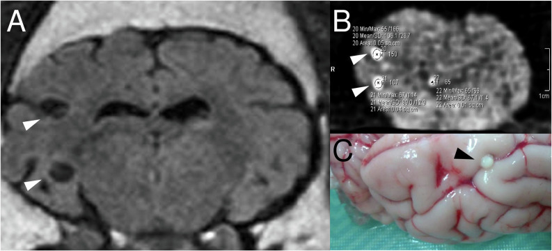
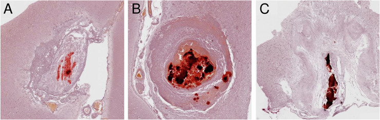
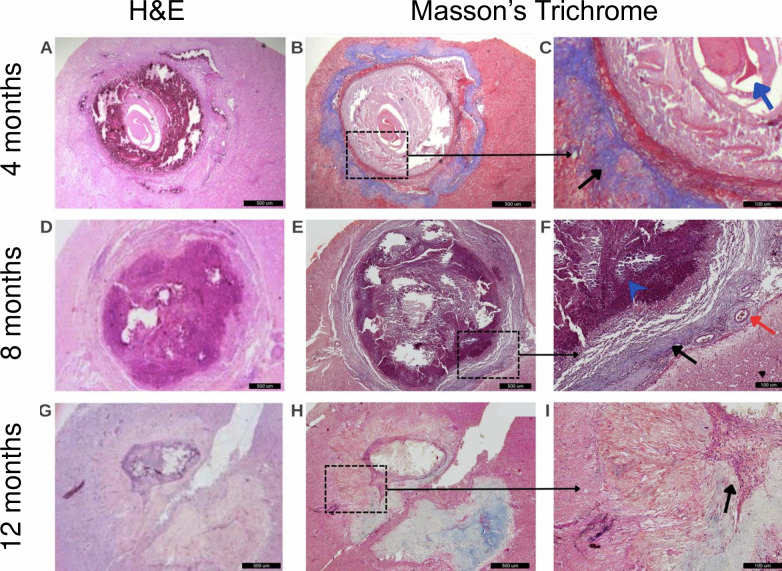
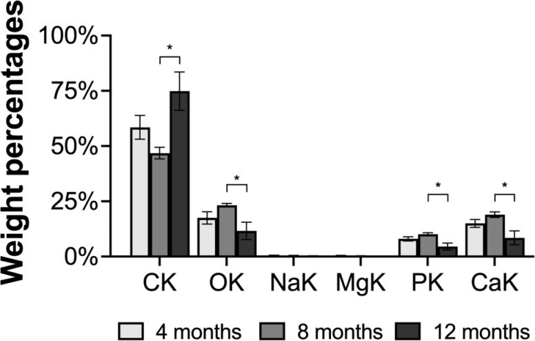
Methods/principal findings: Fifteen naturally infected pigs with viable NCC confirmed by magnetic resonance imaging received APT with albendazole and praziquantel and were sacrificed after 4, 8, and 12 months (n = 5 each). The pigs' brains were removed and processed by ex vivo CT scan to assess the proportion of cysts that calcified by post-treatment time points using risk ratios (RR) from Poisson regression. Radiodensity levels (Hounsefield units) of calcified lesions were also measured and compared using linear coefficients from log-transformed values in generalized linear models. The overall proportion of residual calcification on CT scan was 63.9% (156 calcified lesions/244 viable cysts), being statistically higher in treated NCC pigs at 4 months (83.3% [50/60], RR = 2.61, P < 0.001) and 8 months (82.8% [77/93], RR = 2.59, P < 0.001) versus 12 months (31.9% [29/91]). At 8 months after APT, calcifications were more dense (100.6 ± 3.6 HU) compared to 12 months (74.4 ± 3.6 HU, β = 0.37, P = 0.010) and marginally higher compared to 4 months (85.2 ± 3.8 HU, β = 0.24, P = 0.096), and were also larger and more frequently found on histopathology.
Conclusion/significance: Calcification in NCC is a dynamic process that can be induced and monitored in naturally infected pigs. Eight months after treatment seems to be an optimal time point for assessing residual calcification.
Copyright: © 2025 Arroyo et al. This is an open access article distributed under the terms of the Creative Commons Attribution License, which permits unrestricted use, distribution, and reproduction in any medium, provided the original author and source are credited.
Conflict of interest statement
The authors have declared that no competing interests exist
Figures
References
-
- Garcia HH, Nash TE, Del Brutto OH. Clinical symptoms, diagnosis, and treatment of neurocysticercosis. Lancet Neurol. 2014;13 12:1202-15; doi: 10.1016/S1474-4422(14)70094-8 https://www.ncbi.nlm.nih.gov/pubmed/25453460. - DOI - PMC - PubMed
-
- Ndimubanzi PC, Carabin H, Budke CM, Nguyen H, Qian Y-J, Rainwater E, et al.. A systematic review of the frequency of neurocyticercosis with a focus on people with epilepsy. PLoS Negl Trop Dis. 2010;4(11):e870; doi: 10.1371/journal.pntd.0000870 https://www.ncbi.nlm.nih.gov/pubmed/21072231 - DOI - PMC - PubMed
MeSH terms
Substances
LinkOut - more resources
Full Text Sources
