Effects of fluid shear stress duration on the mechanical properties of HeLa cells using atomic force microscopy
- PMID: 40323916
- PMCID: PMC12052195
- DOI: 10.1371/journal.pone.0321296
Effects of fluid shear stress duration on the mechanical properties of HeLa cells using atomic force microscopy
Abstract
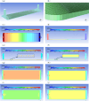
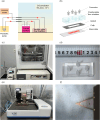
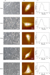
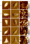
Cellular mechanical properties play a critical role in physiological and pathological processes, with fluid shear stress being a key determinant. Despite its importance, the impact of fluid shear stress on the mechanical characteristics of HeLa cells and its role in the mechanism of tumor metastasis remain poorly understood. This study aims to investigate the effects of varying durations of fluid shear stress on the mechanical properties of HeLa cells, thereby elucidating the mechanical interactions between the fluid flow environment and cancer cells during tumor metastasis. We established an in vitro fluid shear stress cell experimental system and analyzed the flow field characteristics within a parallel plate flow chamber using computational fluid dynamics software. Atomic force microscopy was used to measure the mechanical properties of HeLa cells at different time points under a fluid shear stress of 10 dyn/cm², a value representative of physiological conditions. computational fluid dynamics analysis confirmed the stability of laminar flow and the uniformity of shear stress within the parallel plate flow chamber. The experimental results revealed that with increasing fluid shear stress exposure duration, HeLa cells exhibited a fusiform shape, with a reduction in cell height and a significant decrease in cell Young's modulus. By integrating atomic force microscopy with the in vitro fluid shear stress cell experimental system, this study demonstrates the substantial influence of fluid shear stress on the mechanical properties of HeLa cells. This provides novel insights into the behavior of cancer cells within the in vivo flow environment. Our findings enhance the understanding of cellular mechanical property regulation and offer valuable insights for biomedicine engineering research.
Copyright: © 2025 Zhao et al. This is an open access article distributed under the terms of the Creative Commons Attribution License, which permits unrestricted use, distribution, and reproduction in any medium, provided the original author and source are credited.
Conflict of interest statement
The authors have declared that no competing interests exist.
Figures


References
-
- Ladjal H, Hanus JL, Pillarisetti A, Keefer C, Ferreira A, Desai JP, editors. Atomic force microscopy-based single-cell indentation: Experimentation and finite element simulation. 2009 IEEE/RSJ International Conference on Intelligent Robots and Systems; 2009 10-15 Oct.
-
- Heng YYJQ, Yu W Q. Measurement methods and application of mechanical properties of cells. Sci Sin Vitae. 2023;53(09):1247–73.
MeSH terms
LinkOut - more resources
Full Text Sources

