. 2025 May 13;122(19):e2500356122.
doi: 10.1073/pnas.2500356122. Epub 2025 May 5.

The SIK3-N783Y mutation is associated with the human natural short sleep trait

Affiliations

The SIK3-N783Y mutation is associated with the human natural short sleep trait

Hongmin Chen et al. Proc Natl Acad Sci U S A. .

Abstract

Sleep is an essential component of our daily life. A mutation in human salt induced kinase 3 (hSIK3), which is critical for regulating sleep duration and depth in rodents, is associated with natural short sleep (NSS), a condition characterized by reduced daily sleep duration in human subjects. This NSS hSIK3-N783Y mutation results in diminished kinase activity in vitro. In a mouse model, the presence of the NSS hSIK3-N783Y mutation leads to a decrease in sleep time and an increase in electroencephalogram delta power. At the phosphoproteomic level, the SIK3-N783Y mutation induces substantial changes predominantly at synaptic sites. Bioinformatic analysis has identified several sleep-related kinase alterations triggered by the SIK3-N783Y mutation, including changes in protein kinase A and mitogen-activated protein kinase. These findings underscore the conserved function of SIK3 as a critical gene in human sleep regulation and provide insights into the kinase regulatory network governing sleep.

Keywords: SIK3; kinase; natural short sleep; sleep need.


Conflict of interest statement

Competing interests statement:The authors declare no competing interest.

Figures
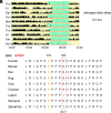
Fig. 1.
Fig. 1.
SIK3-N783Y mutation was identified in a natural short sleep family. (A) Actogram of the subject carrying the SIK3 mutation. (B) SIK3-N783Y is localized to the Proline-rich domain and is conserved among mammals and birds.
Fig. 2.
Fig. 2.
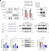
SIK3-N783Y lead to decreased kinase activity. (A) In vitro kinase assay of recombinant SIK3-WT and SIK3-N783Y with recombinant HDAC4/5 as substrates and immunoblot with HDAC4/5 phosphorylation specific antibody. (B) Quantified results for (A). The phosphosites in HDAC4/5 are shown on the Top. (C) In vitro kinase assay with SIK inhibitor YKL-06-062 as control. (D) Schematic of different SIK3 mutations. (E) In vitro kinase assay similar to (A) but with different mutant forms of SIK3. (F) Quantified results for (E). (G) Representative immunoblots with an anti-SPP antibody for different forms of immunoprecipitated SIK3 in the transfected HEK293 cells. (H) Quantified results for (G). *P < 0.05, **P < 0.01, ****P < 0.0001, n.s. not significant. unpaired t test for (B). One-way ANOVA, post hoc Tukey’s multiple comparisons test (F and H). Data are mean ± SEM.
Fig. 3.
Fig. 3.
Sik3-N783Y mice demonstrate reduced sleep time. (AC) Total wake (A), NREM sleep (B), and REM sleep (C) time within 24 h, light phase, and dark phase were measured by EEG/EMG. (D) NREM sleep delta power was plotted hourly over 24 h. (E) Schematic of sleep deprivation (SD) workflow. (FH) NREM (F), REM (G), and total sleep (H) time within 18 h (18 h), light phase (6 to 12 h), and dark phase (12 to 24 h) measured by EEG/EMG were calculated. (I) NREM sleep delta power was plotted hourly over 18 h following SD. Sleep measurements for Sik3+/+ (WT) (n =16), Sik3NY/+(n = 19), Sik3NY/NY (n = 15) under basal conditions. Sleep times for Sik3+/+ (WT) (n = 11), Sik3NY/+ (n = 9) and Sik3NY/NY (n = 13) after SD. *P < 0.05, **P < 0.01. One-way ANOVA (A to C, F to H); two-way ANOVA, post hoc Sidak’s multiple comparisons test (D and I). Data are mean ± SEM.
Fig. 4.
Fig. 4.
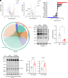
Brains of Sik3N783Y/N783Y mutant (Mut) mice exhibited extensive hypophosphorylation. (A) Experiment workflow for proteomic and phosphoproteomic analysis. (B) Summary of quantified significant change in the phosphoproteome and proteome. (C and D) Volcano plots showing changes of all phosphopeptides in phosphoproteome (C) and proteins in proteome (D). (E and F) Pie chart indicating the fraction of hyperphosphorylated (E) and hypophosphorylated (F) peptides which also exhibited significant increase(red) or decrease (blue) in protein abundance. Light red and light blue indicate alterations exclusively in protein phosphorylation, without corresponding changes in total protein abundance.
Fig. 5.
Fig. 5.
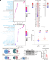
N783Y mutation led to phosphorylation changes at synapse. (A and B) Top20 Gene Ontology (GO) cellular component (CC) (A) and biological process (BP) (B) annotations for the DEPs in Mut/WT comparison. (C) Venn diagram of quantified DEPs (Left) and phosphoproteins (Right) overlapped with previously identified total (Upper) and cycling (Lower) synaptic phosphopeptides and phosphoproteins. (D) Heatmap profile of changes of SNIPPs in the phosphorylation state (Left panel) and total protein abundance (Right panel) in two comparisons. (E) Mean phosphorylation changes of SNIPPs in two animal models. (F) Representative immunoblots using indicated antibodies with the forebrain lysate collected from WT and Mut mice. (G) Quantified results for (F). Mut/WT: Sik3NY/NY vs. WT; Slp/WT: Sik3Sleepy/+ vs. WT. n.s. not significant, **P < 0.01. Chi square test (E), unpaired t test (G).
Fig. 6.
Fig. 6.
N783Y mutation led to phosphorylation changes of kinases. (A and B) Volcano plot showing changes of all putative kinases in phosphoproteome (A) and proteome (B) in the Sik3NY/NY (Mut)/WT group. (C) Enrichment results of kinase–substrate relationships were organized and visualized in a chord plot. The most enriched kinases were shown on the Right (red rings), while the substrates were displayed on the Left (green rings). Every colored chord represents the kinase–substrate relationship originated from one kinase. (D) KSEA enrichment in the Mut/WT group. (E) Representative immunoblots using antibody against phospho-PKA in the forebrain lysate from Sik3WT and Sik3NY/NY mice. (F) Quantified results for (E). (G) Same as (E) except antibody against phospho-MAPK substrate motif was used. (H) Quantified results for (G). *P < 0.05, unpaired t test.

References

    1. Ashbrook L. H., Krystal A. D., Fu Y.-H., Ptáček L. J., Genetics of the human circadian clock and sleep homeostat. Neuropsychopharmacology 45, 45–54 (2020). - PMC - PubMed
    1. Shi G., Wu D., Ptáček L. J., Fu Y.-H., Human genetics and sleep behavior. Curr. Opin. Neurobiol. 44, 43–49 (2017). - PMC - PubMed
    1. Xing L., et al. , Mutant neuropeptide S receptor reduces sleep duration with preserved memory consolidation. Sci. Transl. Med. 11, eaax2014 (2019). - PMC - PubMed
    1. Shi G., et al. , Mutations in metabotropic glutamate receptor 1 contribute to natural short sleep trait. Curr. Biol. 31, 13–24.e4 (2021). - PMC - PubMed
    1. Shi G., et al. , A rare mutation of β1-adrenergic receptor affects sleep/wake behaviors. Neuron 103, 1044–1055.e7 (2019). - PMC - PubMed

LinkOut - more resources