

Acute epididymo-orchitis complicated by outcomes of either testicular necrosis or complete recovery: Two case reports
- PMID: 40324246
- PMCID: PMC12055044
- DOI: 10.1097/MD.0000000000042391
Acute epididymo-orchitis complicated by outcomes of either testicular necrosis or complete recovery: Two case reports
Abstract
Rationale: Acute epididymo-orchitis, a common urological emergency requiring prompt intervention to prevent complications like testicular ischemia. This study highlights the use of serial Doppler ultrasound monitoring in patients with acute epididymo-orchitis, particularly in high-risk individuals.
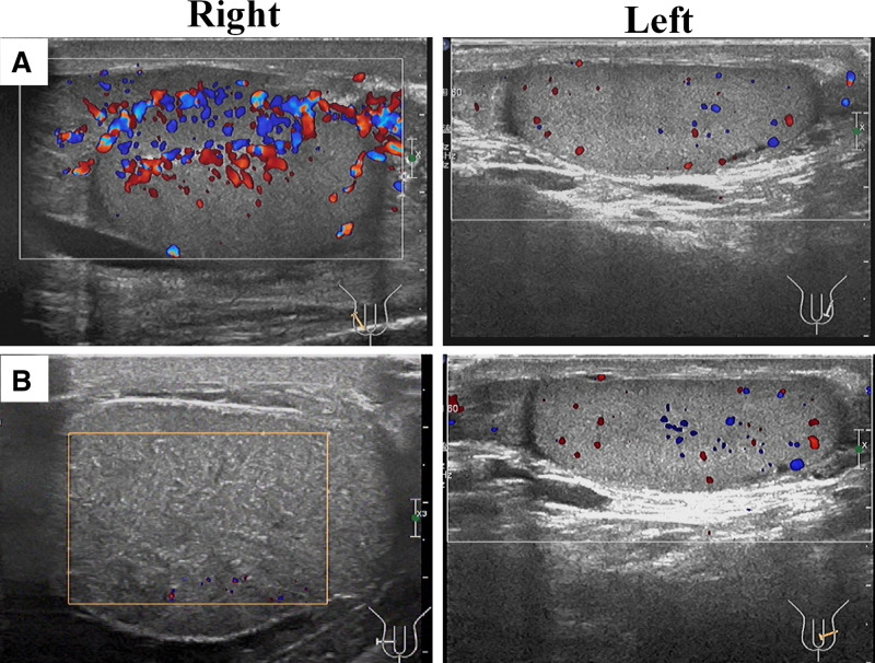
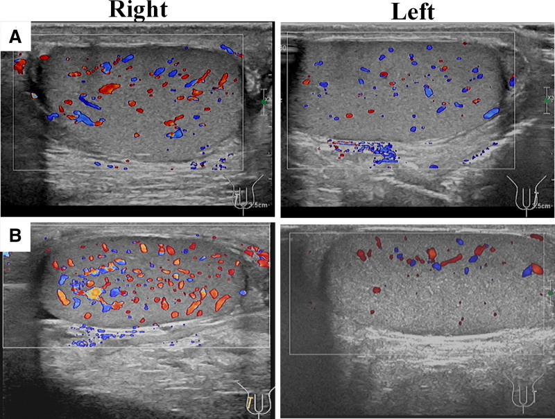
Patient concerns: Case 1: A 70-year-old male with a history of bladder cancer, prostate malignancy, and hypertension, presented with scrotal swelling, pain, and lower abdominal discomfort. Physical examination revealed an enlarged and tender right testicular epididymis, with normal findings on the left. Ultrasound showed increased blood flow to the right testicular epididymis, indicating inflammation. Case 2: A 34-year-old male presented with scrotal swelling, pain, and lower abdominal discomfort. Ultrasound revealed increased testicular and epididymal blood flow, suggesting inflammation. The antibiotic therapy was adjusted according to the continuous ultrasound monitoring.
Diagnoses: Both cases were ultimately diagnosed as testicular epididymal inflammation.
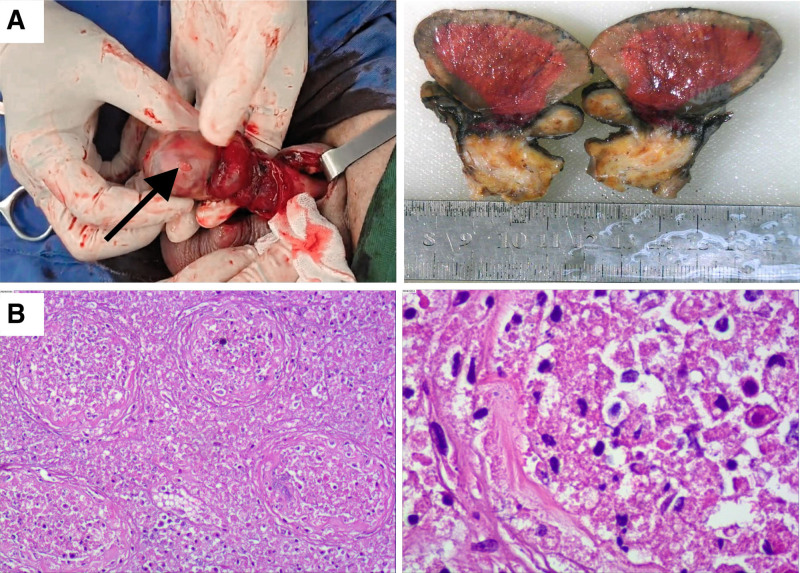
Interventions: Patient 1 underwent anti-inflammatory therapy and orchiectomy. Patient 2 was treated with antibiotic therapy and recovered.
Outcomes: Patient 1 experienced testicular necrosis, whereas Patient 2 achieved a full recovery.
Lessons: The importance of serial Doppler ultrasonography: delayed follow-up imaging in this case allowed ischemic changes to progress irreversibly, despite initial Doppler findings showing increased perfusion. Early and repeated imaging is critical to monitor disease progression and guide timely interventions. Limitations of inflammatory markers: the disease worsened although the patient's leukocytosis and IL-6 levels improved markedly during treatment, highlighting that relying solely on blood tests is insufficient to determine treatment efficacy. Patient-specific risk stratification: high-risk individuals require more aggressive diagnostic and therapeutic protocols to prevent irreversible complications.
Keywords: case report; complication; epididymo-orchitis; orchiectomy; testicular necrosis.
Copyright © 2025 the Author(s). Published by Wolters Kluwer Health, Inc.
Conflict of interest statement
The authors have no conflicts of interest to disclose.
Figures
Similar articles
-
Multimodal ultrasound diagnosis of epididymo-orchitis with secondary testicular infarction: A case report.J Clin Ultrasound. 2024 Jul-Aug;52(6):813-819. doi: 10.1002/jcu.23692. Epub 2024 Apr 16. J Clin Ultrasound. 2024. PMID: 38624174
-
Idiopathic testicular infarction in a boy initially suspected to have acute epididymo-orchitis associated with mycoplasma infection and Henoch-Schönlein purpura.J Pediatr Urol. 2009 Feb;5(1):68-71. doi: 10.1016/j.jpurol.2008.07.003. Epub 2008 Aug 26. J Pediatr Urol. 2009. PMID: 18753011
-
Acute Epididymo-orchitis-Related Global Testicular Infarction: Clinical and Ultrasound Findings With an Emphasis on the Juxta-epididymal String-of-Bead Sign.Ultrasound Q. 2016 Sep;32(3):283-9. doi: 10.1097/RUQ.0000000000000225. Ultrasound Q. 2016. PMID: 27556195
-
Challenges in the diagnosis of testicular infarction in the presence of prolonged epididymitis: Three cases report and literature review.J Xray Sci Technol. 2020;28(4):809-819. doi: 10.3233/XST-200671. J Xray Sci Technol. 2020. PMID: 32474478 Review.
-
Brucellar epididymo-orchitis: a retrospective multicenter study of 28 cases and review of the literature.Travel Med Infect Dis. 2014 Nov-Dec;12(6 Pt A):667-72. doi: 10.1016/j.tmaid.2014.10.005. Epub 2014 Oct 25. Travel Med Infect Dis. 2014. PMID: 25457303 Review.
References
-
- Sedrak M, Song A, Greenstein J, Hahn B. Man with scrotal pain. Ann Emerg Med. 2023;82:111–4. - PubMed
-
- Medina-Polo J. Treatment of Epididymitis and Orchitis[M]//Guide to Antibiotics in Urology. Springer International Publishing; 2024:229–238.
-
- Ramírez-González J A, Sansone A. Male Reproductive System [M]// Fertility, Pregnancy, and Wellness. Elsevier; 2022:23–36.
Publication types
MeSH terms
Substances
Grants and funding
LinkOut - more resources
Full Text Sources
