. 2025 Nov;83(5):1077-1091.
doi: 10.1016/j.jhep.2025.04.034. Epub 2025 May 3.

New platinum derivatives selectively cause double-strand DNA breaks and death in naïve and cisplatin-resistant cholangiocarcinomas

Affiliations

New platinum derivatives selectively cause double-strand DNA breaks and death in naïve and cisplatin-resistant cholangiocarcinomas

Irene Olaizola et al. J Hepatol. 2025 Nov.

Abstract

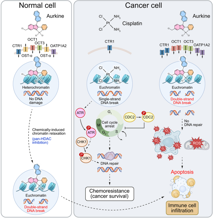
Background & aims: Patients with cholangiocarcinoma (CCA) have poor prognosis. Current cisplatin-based first-line chemotherapy offers limited survival benefit. Cisplatin induces single-strand DNA breaks, activating DNA repair mechanisms that diminish its effectiveness. Here, we present the design, chemical synthesis, and therapeutic evaluation of a new generation of chemotherapeutic agents (Aurkines) with unique polyelectrophilic properties. These agents cause a high frequency of double-strand DNA breaks, bypassing DNA repair, and promoting cancer cell death.

Methods: Two novel compounds, Aurkine 16 and Aurkine 18, were designed and evaluated for their antitumor effects in both naïve and cisplatin-resistant CCA cells, cancer-associated fibroblasts, healthy cholangiocytes, and in vivo models.

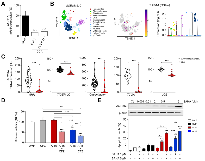
Results: Aurkines effectively induced double-strand DNA breaks, leading to increased DNA damage and elevated levels of reactive oxygen species, resulting in greater cytotoxicity than cisplatin in CCA cells. Phosphoproteomic and molecular analysis revealed that cisplatin activates DNA repair pathways, while Aurkines primarily induce apoptosis. Importantly, Aurkines also triggered apoptosis in cisplatin-resistant CCA cells and cancer-associated fibroblasts without harming healthy cholangiocytes. Additionally, Aurkines demonstrated cytotoxicity in other cisplatin-resistant cancers, such as breast and ovarian cancer. This tumor selectivity results from reduced uptake, increased efflux, and compact chromatin structure in normal cells, limiting Aurkine-DNA interactions. In vivo, Aurkines inhibited the growth of subcutaneous naïve and cisplatin-resistant CCA tumors, as well as orthotopic tumors in immunocompetent mice, promoting antitumor immune cell recruitment without any adverse events. Transport studies revealed that Aurkines were selectively taken up by OCT1, OCT3, CTR1, and OATP1A2, whereas only CTR1 transported cisplatin.

Conclusions: Aurkines represent promising therapeutic drugs for both naïve and cisplatin-resistant cancers due to their unique polyelectrophilic properties and selective targeting of malignant cells.

Impact and implications: This study introduces a novel therapeutic strategy designed to induce frequent double-strand DNA breaks selectively in both naïve and cisplatin-resistant cancer cells, without evident toxic side effects at therapeutic doses. This approach may form the basis for new strategies to overcome the critical challenge of drug resistance in cancer treatment and has the potential to be a breakthrough not only for the treatment of biliary tumors but also for other cancers.

Keywords: Cancer; Chemoresistance; Chemotherapy; DNA damage.


Conflict of interest statement

Conflict of interest FPC is scientific advisor of Quimatryx Ltd (quimatryx.com). Remaining authors have no conflicts of interest to declare related to this manuscript. Please refer to the accompanying ICMJE disclosure forms for further details.

Figures

Image 1
Graphical abstract
Fig. 1
Fig. 1
Design, structure, and interaction modes of Aurkines. (A) Interaction of dielectrophilic (E2) and trielectrophilic (E3) platinum reagents with guanine (G) units. (B) Chemical structures of Aurkines 16 and 18. (C) Molecular dynamics simulations illustrating the interaction of Aurkine 16 with DNA, highlighting both the initial minor groove binding associated with the β-naphthyl moiety (pale blue) and the subsequent major groove intercalation of the heterocyclic groups (pink and orange).
Fig. 2
Fig. 2
Effect of Aurkine on isolated DNA. (A) Atomic force microscopy (AFM) images of untreated DNA and DNA incubated with CisPt or Aurkine 16 for 10 min. (B) Transmission electron microscopy (TEM) images of untreated DNA, and DNA incubated with Aurkine 16 for 10 min. (C) Agarose gel analysis of pUC18 plasmid after 1 h incubation with vehicle, CisPt, Aurkine 16 or 18. HindIII digestion was used as control. (D) Venn diagram showing the number of canonical pathways with significant adjusted p values in both comparisons and a heatmap illustrating the altered protein phosphorylation in EGI-1 cells after a 3-hour exposure to CisPt or Aurkines. Enriched proteins are coloured in red and proteins with lower abundance are displayed in blue. CisPt, cisplatin; DMF, dimethylformamide; SC, supercoiled.
Fig. 3
Fig. 3
Genotoxic effects of Aurkines 16 and 18 on human CCA cells. (A) DNA damage, (B) ROS and (C) mROS in NHCs and CCA cell lines (HUCCT1 and EGI-1) after 24-hour incubation with vehicle, CisPt, Aurkines 16 or 18 (10 μM). (D) Cell viability of NHCs and CCA cell lines (HUCCT1 and EGI-1) after 48-hour incubation with vehicle, CisPt, Aurkines 16 or 18 (10 μM and 20 μM). One-way ANOVA test or Student’s t tests were used. Data are shown as mean ± SEM. ∗p ≤0.05, ∗∗p ≤0.01, ∗∗∗p ≤0.001). CCA, cholangiocarcinoma; CisPt, cisplatin; DMF, dimethylformamide; NHCs, normal human cholangiocytes; mROS, mitochondrial ROS; ROS, reactive oxygen species.
Fig. 4
Fig. 4
Pro-apoptotic effects of Aurkines 16 and 18 on human CCA cells. (A) % of cleaved caspase-3+ cells and (B) Annexin V/TO-PRO™-3 dual staining of NHCs and CCA cell lines (HUCCT1 and EGI-1) after 48-hour incubation with vehicle, CisPt, Aurkines 16 or 18 (10 μM and 20 μM). (C) Cell proliferation and (D) cell cycle analysis in NHCs and CCA cell lines (HUCCT1 and EGI-1) after 24-hour incubation with vehicle, CisPt, Aurkines 16 or 18 (10 μM). One-way ANOVA test or Student’s t tests were used. Data are shown as mean ± SEM. ∗p ≤0.05, ∗∗p ≤0.01, ∗∗∗p ≤0.001. CCA, cholangiocarcinoma; CisPt, cisplatin; DMF, dimethylformamide; NHCs, normal human cholangiocytes.
Fig. 5
Fig. 5
Mechanisms of NHC resistance to Aurkines. (A) SLC51A mRNA expression in NHC and CCA cell lines (HUCCT1 and EGI-1). (B) t-SNE plot clustering all the cell populations detected by scRNA sequencing in the GSE151530 dataset, which includes samples from 12 patients with CCA. (C) SLC51A mRNA expression levels in CCA tumors compared to SL tissue from the AHN, TIGER-LC, Copenhagen, TCGA and JOB human cohorts. (D) NHC viability after 48-hour incubation with Aurkines 16 and 18 (20 μM), or their combination with the OST-α/β inhibitor (CFZ, 10 μM). (E) Immunoblot of Ac–H3K9 in NHCs after 48-hour incubation with SAHA, with β-actin as loading control. Apoptosis of NHCs after 48-hour incubation with CisPt, Aurkines 16 and 18 (10 μM), or their combination with SAHA (1 μM and 5 μM). One-way ANOVA test or Student’s t tests were used. Data are shown as mean ± SEM. ∗p ≤0.05, ∗∗p ≤0.01, ∗∗∗p ≤0.001. CCA, cholangiocarcinoma; CFZ, clofazimine; CisPt, cisplatin; DMF, dimethylformamide; NHCs, normal human cholangiocytes; OST, organic solute transporter; SAHA, suberoylanilide hydroxamic acid; SL, surrounding liver.
Fig. 6
Fig. 6
Antitumor effect of Aurkine 16 and 18 on CisPt-resistant CCA cells (EGI-1R) and CAFs. (A) Apoptosis in EGI-1 and EGI-1R cells after 48-hour incubation with increasing CisPt doses. (B) DNA damage, (C) ROS and (D) mROS in EGI-1R cells after 24-hour incubation with vehicle, CisPt, Aurkines 16 or 18 (10 μM). (E) Cell viability, (F) % of cleaved caspase-3+ cells and (G) Annexin V/TO-PRO™-3 dual staining of EGI-1R cells after 48-hour incubation with vehicle, CisPt, Aurkines 16 or 18 (10 μM and 20 μM). (H) Cell proliferation and (I) cell cycle analysis in EGI-1R cells after 24-hour incubation with vehicle, CisPt, Aurkines 16 or 18 (10 μM). (J) Cell viability and apoptosis of CAFs after 48-hour incubation with vehicle, CisPt, Aurkines 16 or 18 (10 and 20 μM). One-way ANOVA test or Student’s t tests were used. Data are shown as mean ± SEM. ∗p ≤0.05, ∗∗p ≤0.01, ∗∗∗p ≤0.001. CAFs, cancer-associated fibroblasts; CCA, cholangiocarcinoma; CisPt, cisplatin; DMF, dimethylformamide; mROS, mitochondrial ROS; ROS, reactive oxygen species.
Fig. 7
Fig. 7
In vivo antitumor activity of Aurkines 16 and 18 in experimental murine models of CCA. (A) Schematic representation of the subcutaneous CCA model. (B) Tumor volume growth during treatment with CisPt or Aurkine 16 (0.5 mg/kg) in the subcutaneous CCA model. Group sizes: vehicle control (n = 12), CisPt (n = 11), Aurkine 16 (n = 13). (C) Representative tumor images, along with Ki67 and cleaved caspase-3 staining images and quantification in the subcutaneous CCA model. (D) Schematic representation of the subcutaneous CisPt-resistant CCA model. (E) Tumor volume growth during treatment with CisPt, Aurkines 16 and 18 (2 mg/kg) in the subcutaneous CisPt-resistant CCA model. Group sizes: vehicle control (n = 13), CisPt (n = 10), Aurkine 16 (n = 10), Aurkine 18 (n = 16). (F) Schematic representation of the orthotopic CCA model. (G) Representative macroscopic images of liver tumors from vehicle-, CisPt-, and Aurkine 16-treated animals. (H) Tumor volume at sacrifice following treatment with CisPt and Aurkine 16 (0.5 mg/kg) in the orthotopic CCA model. Group sizes: vehicle control (n = 16), CisPt (n = 16), Aurkine 16 (n = 16). (I) Representative images and quantification of p-H2AX staining in the orthotopic CCA model. (J) Representative images and quantification of CD4 and CD8 staining in the orthotopic CCA model. One-way ANOVA test or Student’s t test were used. Data are shown as mean ± SEM. p values (B, E): #p <0.05, compared to vehicle-treated; ∗p <0.05, compared to CisPt-treated. p values (C, H-J): ∗p ≤0.05, ∗∗p ≤0.01, ∗∗∗p ≤0.001. Scale bar: 100 μm. CCA, cholangiocarcinoma; CisPt, cisplatin; DMF, dimethylformamide.
Fig. 8
Fig. 8
Analysis of transporters involved in the uptake of Aurkines 16 and 18 by cancer cells. (A) Uptake of specific fluorescent substrates by cells with or without experimental overexpression of each transporter, measured by flow cytometry. (B) Intracellular accumulation of Aurkine 16 and 18 in cells with or without overexpression of each transporter, measured by HPLC-MS/MS. (C) Relative mRNA expression (qPCR) of SLC22A3 in NHCs, CAFs, eCCA (i.e. EGI-1, EGI-1R, TFK1, WITT) and iCCA (i.e. HUCCT1, RBE and SNU-1079) cells. (D) Expression levels of SLC22A3 at the single-cell level in human CCA tumors. (E) mRNA expression of SLC22A3 in CCA tissues, stratified by mutational status (IDH1, KRAS, TP53 or WT). (F) mRNA expression of SLC22A3 in CCA tissues, stratified by anatomical origin (iCCA vs. eCCA). (G) mRNA expression of SLC22A3 in CCA cell lines, stratified by anatomical origin (iCCA vs. eCCA). One-way ANOVA test or Student’s t tests were used. Data are shown as mean ± SEM. ∗p ≤0.05, ∗∗p ≤0.01, ∗∗∗p ≤0.001. CisPt, cisplatin; CTR, copper transporter; DCs, dendritic cells; DMF, dimethylformamide; eCCA, extrahepatic cholangiocarcinoma; iCCA, intrahepatic cholangiocarcinoma; NHCs, normal human cholangiocytes; OATP, organic anion transporting polypeptide; OCT, organic cation transporter; Qui, quinine; Rif, rifampicine; WT, wild-type. This figure was partly generated using Servier Medical Art, provided by Servier, licensed under a Creative Commons Attribution 3.0 unported license.

References

    1. Banales J.M., Marin J.J.G., Lamarca A., et al. Cholangiocarcinoma 2020: the next horizon in mechanisms and management. Nat Rev Gastroenterol Hepatol. 2020;17:557–588. - PMC - PubMed
    1. Izquierdo-Sanchez L., Lamarca A., La Casta A., et al. Cholangiocarcinoma landscape in Europe: diagnostic, prognostic and therapeutic insights from the ENSCCA Registry. J Hepatol. 2022;76:1109–1121. - PubMed
    1. Valle J., Wasan H., Palmer D.H., et al. Cisplatin plus gemcitabine versus gemcitabine for biliary tract cancer. N Engl J Med. 2010;362:1273–1281. - PubMed
    1. Oh D.-Y., Ruth He A., Qin S., et al. Durvalumab plus gemcitabine and cisplatin in advanced biliary tract cancer. NEJM Evid. 2022;1:1–11. - PubMed
    1. Kelley R.K., Ueno M., Yoo C., et al. Pembrolizumab in combination with gemcitabine and cisplatin compared with gemcitabine and cisplatin alone for patients with advanced biliary tract cancer (KEYNOTE-966): a randomised, double-blind, placebo-controlled, phase 3 trial. The Lancet. 2023;401:1853–1865. - PubMed

MeSH terms