. 2025 May 5;16(1):4168.
doi: 10.1038/s41467-025-59538-z.

Nageotte nodules in human dorsal root ganglia reveal neurodegeneration in diabetic peripheral neuropathy

Affiliations

Nageotte nodules in human dorsal root ganglia reveal neurodegeneration in diabetic peripheral neuropathy

Stephanie I Shiers et al. Nat Commun. .

Abstract

Nageotte nodules, first described in 1922 by Jean Nageotte, are clusters of non-neuronal cells that form after sensory neuron death. Despite their historical recognition, little is known about their molecular identity nor their involvement in neuropathies that involve neuronal loss like diabetic peripheral neuropathy (DPN). In this study, we molecularly characterize Nageotte nodules in dorsal root ganglia recovered from organ donors with DPN. Here we show that Nageotte nodules are abundant in DPN sensory ganglia and account for 25% of all neurons. Peripherin-and Nav1.7-positive dystrophic axons invade Nageotte nodules, forming small neuroma-like structures. Using histology and spatial sequencing, we demonstrate that Nageotte nodules are mainly composed of satellite glia and non-myelinating Schwann cells that express SPP1 and are intertwined with sprouting sensory axons originating from neighboring neurons. Our findings suggest that Nageotte nodules are an integral feature of dorsal root ganglion neurodegeneration, providing potential therapeutic targets for sensory neuron protection and pain management in DPN.


Conflict of interest statement

Competing interests: T.J.P. is a co-founder of 4E Therapeutics. The authors declare no other competing interests related to this work.

Figures

Fig. 1
Fig. 1. Identification of Nageotte nodules in DRGs from diabetic organ donors.
A Hematoxylin and eosin staining was performed on DRGs from organ donors (n = 90), B and then each DRG was scored for prevalence of Nageotte nodules using a qualitative scoring system. C After scoring, donors were grouped based on their medical history of diabetes, analgesic usage, or medical note of diabetic peripheral neuropathy (DPN). Diabetic donors had significantly higher Nageotte nodule scores compared to non-diabetic donors without pre-existing pain conditions. In diabetics, Nageotte nodule content increased in severity in relation to DPN as indicated by analgesic usage, medical note of DPN, and/or diabetes-related amputation. D Representative images of an L4 bi-ganglia from a DPN donor immunostained for GFAP (red, satellite glial cells and non-myelinating Schwann cells), peripherin (green, sensory neurons), and DAPI (blue, nuclei). Asterisks denote Nageotte nodules. Sample size: Non-diabetic n = 7; DPN n = 9. E The percentage of Nageotte nodules was significantly higher in the DPN DRGs (average: 25%) compared to non-diabetic DRGs (average: 8%). F Confocal image of a peripherin-positive axon bundle intertwined with other cells at a Nageotte nodules. Similar staining was observed across all 9 DPN donors. G Image taken from Jean Nageotte’s original 1922 publication in which three Nageotte nodules (mid top, mid bottom, and left) contain axon bundles which sprout from a glomerulus (middle). H) Transmission electron microscopy (TEM) of a Nageotte nodule. Arrows point to unmyelinated axonal fibers. Sample size: Diabetic taking analgesics n = 2. Statistical tests: (C): One-way ANOVA with Bonferroni’s multiple comparisons test. *p = 0.0309, ****p < 0.0001, ns (not-significant) p = 0.5525. E: Unpaired two-sided t-test. **p = 0.0013. Data points represent individual donors. Error bars = mean +/- SEM. Scale bars: (A): 500 μm, 100 μm. 20 μm (B): 100 μm. (D): Mosaic—1 mm and other panels—50 μm. (F): 10 μm. (H): 2 μm.
Fig. 2
Fig. 2. Nageotte nodule axons express nociceptive, but not sympathetic markers.
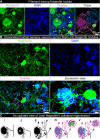
A Tyrosine hydroxylase (TH, red) labeling in combination with peripherin (green) and DAPI (blue) revealed that Nageotte nodule axonal sprouts were not sympathetic in origin. Sample size: DPN n = 9. B TrpV1 fibers at a Nageotte nodule in a DPN DRG (white arrow). TrpV1 was only detected in Nageotte nodules from a single DPN donor out of 5 that were investigated. The TrpV1+ fibers at the Nageotte nodule appeared to arise from a glomerulus (magenta arrow). Sample size: DPN n = 5. C Nav1.7 (red) was detected in the axonal fibers intertwined with Nageotte nodules. Sample size: DPN n = 6. Scale bars: A: 10 μm. B: 20 μm. C: 10 μm.
Fig. 3
Fig. 3. Nageotte nodule axon bundles originate from axonal sprouts from local sensory neurons in situ and in vitro.
A Nageotte nodule (white asterisks), two sensory neuron cell bodies (yellow asterisks), and dystrophic axons (magenta arrows) in a 60X z-stack projection image of a DRG section stained for peripherin (green) and DAPI (blue) from donor 6. In the Trace panel, axonal filaments stemming from the neuronal soma of the middle sensory neuron were traced in Imaris (pink filament trace) which mainly connected to dystrophic axons (multi-colored axonal blebs). The Nageotte nodule axonal bundle (blue filament trace) was mainly spooling fibers that traced back to dystrophic axons. In some cases, dystrophic axons were interconnected to one another (yellow filament traces). Sample size: DPN with highest Nageotte score n = 1. B A representative 20X z-stack projection image of human DRG sensory neurons that were cultured in vitro for 3 days and then stained for peripherin (green) and DAPI (blue). Sample size: Non-diabetic n = 2. Human sensory neurons display a multipolar phenotype in which multiple axonal branches sprout from the neuronal soma (yellow arrows), form dystrophic axons (magenta arrows), and intertwine with structures resembling Nageotte nodules (white arrows). A digitally zoomed-in view of the outlined area (cyan) exemplifies a multipolar sensory neuron sprouting fibers into a Nageotte nodule. C Jean Nageotte described collateral sprouting in 1922 (I–III; original illustration by Jean Nageotte, 1922) in which I) a normal ganglion cell with a T-bifurcated axon is II) deprived of the radicular branch of the axon resulting in III) neurite outgrowth from the soma and glomerulus which are equipped with encapsulated growth balls. Jean Nageotte as well as our imaging of DPN DRGs noted IV) the formation of non-sympathetic pericellular nests that formed around sensory neurons with intact somata and those with shrunken/misshapen somata likely in the process of dying. V) Neurites sprout from the dystrophic axons forming arborizations at Nageotte nodules. Scale bars: A: 15 μm. B: 50 μm and zoomed-in view panel: 10 μm.
Fig. 4
Fig. 4. Spatial transcriptomics of Nageotte nodules identifies non-myelinating Schwann cells and satellite glia as prominent cell types.
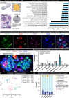
A VISIUM spatial transcriptomics was conducted on 16 DRG sections from 6 DPN donors. Barcodes touching Nageotte nodules (1094) and nearby neurons (1087) were selected for downstream analysis. B) Key gene ontology themes were related to neurodegeneration. C RNAscope in situ hybridization for SOX10 (green, satellite glia and Schwann cells), FABP7 (red, satellite glia), CD68 (purple, macrophages), and DAPI (blue) in a DPN DRG. Confocal, 40X. Sample size: DPN n = 6. D Digitally zoomed overlay images of Nageotte nodule 1 (cyan arrow in (C)) and Nageotte nodule 2 (yellow arrow in (C)). Nageotte nodule 1 had higher content of FABP7+ nuclei, while Nageotte nodule 2 had little-to-no FABP7 signal, indicating that there are differences in the composition of cell types between Nageotte nodules. E Deconvolution using single-nuclear sequencing datasets revealed that the potential sources of the majority of the non-neuronal mRNA transcripts in Nageotte nodules come from Satellite glial cells and non-myelinating (NM) Schwann cells. The remaining percentage of transcripts in Nageotte nodules arise from neurons, likely axonally trafficked mRNAs. Data points are not shown on graph as they represent individual barcodes (n = 1041, 1064, and 39191 for nodules, nearby neurons, and other barcodes, respectively). F Clustering analysis of Nageotte nodule barcodes identified 5 subclusters. G Deconvolution reveals differences in the distribution of estimated mRNA sources between clusters. Statistical tests: B: Fisher’s exact test with correction for multiple comparisons run in Enrichr. Adjusted p-values shown. E: Error bars = mean +/- SD. Scale bars: C: 50 μm. D: 10 μm. Graphic in (A): Created in BioRender. Shiers, S. (2025) https://BioRender.com/5u9g7kp. Source data are provided as a Source Data file.
Fig. 5
Fig. 5. Ligand-receptor interactions between Nageotte nodules and nearby neurons identifies osteopontin (SPP1) and CD44.
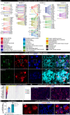
A Differentially expressed ligands per Nageotte nodule cluster and corresponding receptors expressed in nearby neurons. B RNAscope in situhybridization for SCN10A (green, Nav1.8), SPP1 (red, osteopontin), CD44 (blue), and DAPI (cyan) in a diabetic peripheral neuropathy (DPN) DRG. White outline denotes the digitally zoomed-in image of a single Nageotte nodule shown in the bottom panel. Confocal, 40X. Sample size: DPN n = 5. C Top 20 expressed cytokines in Nageotte nodules clusters, showing log10(counts per million). Data points represent individual Nageotte nodule barcodes color coded by cluster (n = 1094). D Immunohistochemistry for phosphorylated eukaryotic translation initiation factor (red, p-eIF4E) and DAPI (blue) in a non-diabetic and DPN DRG. E p-eIF4E was significantly elevated in the soma of sensory neurons in the DPN DRGs. Data points represent individual donors. Sample size: Non-diabetic n = 5, DPN n = 5. F p-eIF4E was also detected at Nageotte nodules in the DPN DRGs. Statistical tests: E: Unpaired two-sided t-test. ***p = 0.0002. C, E: Error bars = mean +/- SEM. Scale bars: B: top panel—50 μm and bottom panel—10 μm. D: 200 μm. F: 50 μm. Source data is provided as a Source Data file.

Update of

References

    1. Nageotte, J. L’organisation de la matiere dans ses rapports avec la vie: etudes d’anatomie generale et de morphologie experimentale sur le tissu conjonctif et le nerf. (F. Alcan, 1922).
    1. Lakritz, J. R. et al. An oral form of methylglyoxal-bis-guanylhydrazone reduces monocyte activation and traffic to the dorsal root ganglia in a primate model of HIV-peripheral neuropathy. J. Neurovirol.23, 568–576 (2017). - PMC - PubMed
    1. Lakritz, J. R. et al. α4-integrin antibody treatment blocks monocyte/macrophage traffic to, vascular cell adhesion molecule-1 expression in, and pathology of the dorsal root ganglia in an SIV macaque model of HIV-peripheral neuropathy. Am. J. Pathol.186, 1754–1761 (2016). - PMC - PubMed
    1. Burdo, T. H., Orzechowski, K., Knight, H. L., Miller, A. D. & Williams, K. Dorsal root ganglia damage in SIV-infected rhesus macaques: an animal model of HIV-induced sensory neuropathy. Am. J. Pathol.180, 1362–1369 (2012). - PMC - PubMed
    1. Lakritz, J. R. et al. Monocyte traffic, dorsal root ganglion histopathology, and loss of intraepidermal nerve fiber density in SIV peripheral neuropathy. Am. J. Pathol.185, 1912–1923 (2015). - PMC - PubMed