. 2025 Sep;62(9):11834-11847.
doi: 10.1007/s12035-025-05005-1. Epub 2025 May 6.

ERRα Knockout Promotes M2 Microglial Polarization and Inhibits Ferroptosis in Sepsis-Associated Brain Dysfunction

Affiliations

ERRα Knockout Promotes M2 Microglial Polarization and Inhibits Ferroptosis in Sepsis-Associated Brain Dysfunction

Jun Jin et al. Mol Neurobiol. 2025 Sep.

Abstract

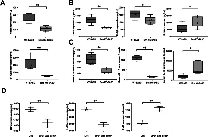
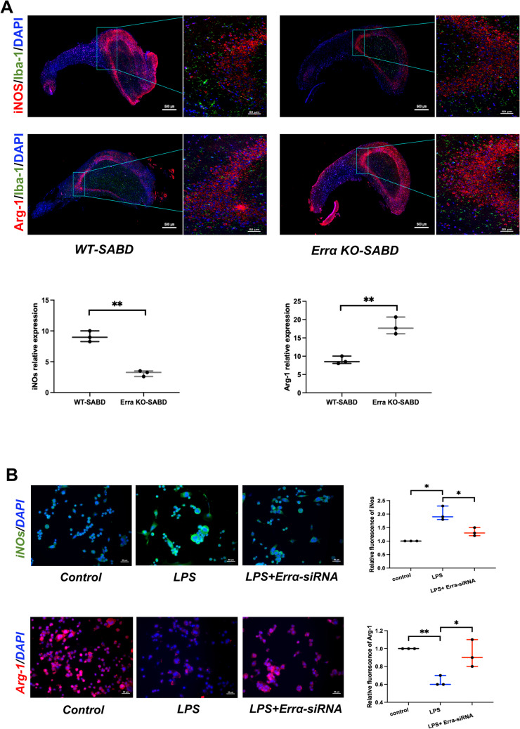
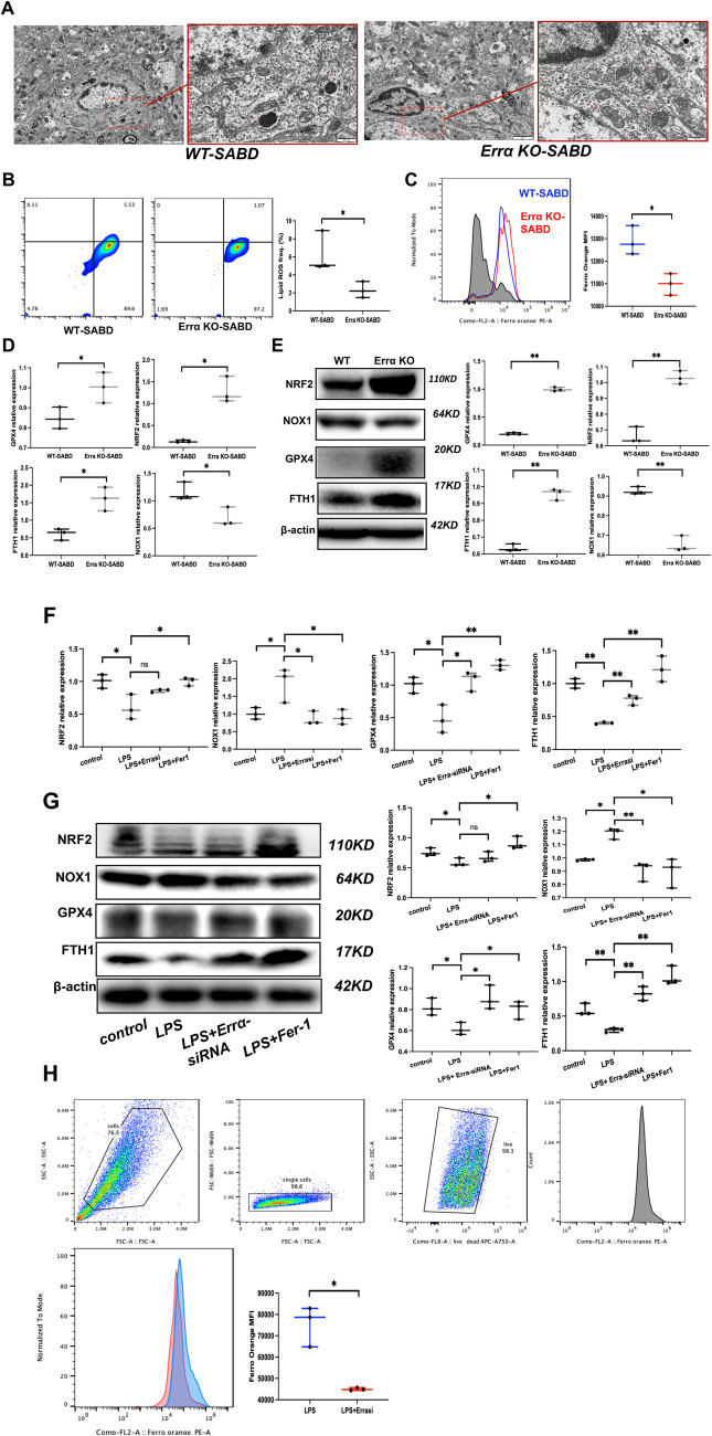
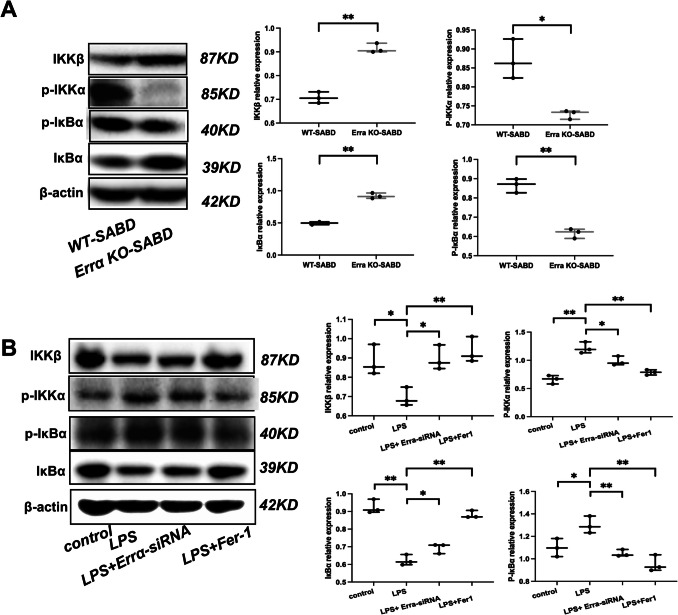
Sepsis-associated brain dysfunction (SABD) is a critical neurological complication with high mortality, yet its pathogenesis remains poorly understood. This study investigated the role of estrogen-related receptor α (ERRα) in SABD pathogenesis using ERRα knockout (KO) mice and cecal ligation and puncture (CLP) models. We found that ERRα KO mice exhibited improved survival rates, milder neurological symptoms, reduced pro-inflammatory cytokine production (TNF-α, IL-1β), and increased anti-inflammatory cytokine (IL-10) levels compared to wild-type controls. Additionally, ERRα deficiency promoted microglial M2 polarization and attenuated ferroptosis, as evidenced by decreased iron accumulation, reduced lipid peroxidation, and normalized mitochondrial morphology. Mechanistically, these protective effects were mediated through inhibition of the NF-κB signaling pathway. In vitro studies with ERRα knockdown in LPS-stimulated BV2 microglia confirmed these findings. Our results suggest that ERRα as a critical regulator of microglial function in SABD through coordinated control of inflammatory responses, polarization states, and ferroptosis, suggesting that targeting ERRα may represent a promising therapeutic strategy for SABD treatment.

Keywords: Estrogen-related receptor α (Errα); Ferroptosis; Inflammatory response; Microglia; NF-κB pathway; Sepsis-associated brain dysfunction.


Conflict of interest statement

Declarations. Ethics Approval and Consent to Participate: This study was approved by the Medical Research Ethics Committee of The University of Hong Kong-Shenzhen Hospital (Ethic-HKUSZH2022062). Consent for Publication: Not applicable. Conflict of interest: The authors declare no competing interests.

Figures

Fig. 1
Fig. 1
Errα KO mouse has milder symptoms and longer survival with sepsis-associated brain dysfunction (SABD). A ERRα knockout transgenic mice were established. B Sepsis-associated brain dysfunction (SABD) mice were established. C The Kaplan–Meier curves for 48-h survival between ERRα KO-SABD and WT-SABD. D H&E staining of the mouse hippocampus (N = 5)
Fig. 2
Fig. 2
Errα-KO alleviated neuroinflammation in Sepsis-associated brain dysfunction (SABD). A NSE and S100 β levels in ErrαKO-SABD and WT-SABD mice determined by ELISA. Data are expressed as mean ± SD (N = 5) **p < 0.01. B TNFα, IL-1β and IL-10 levels in ErrαKO-SABD and WT-SABD mice determined by ELISA. Data are expressed as mean ± SD (N = 5). *p < 0.05, **p < 0.01. C Serum TNFα, IL-1β, and IL-10 levels in ErrαKO-SABD and WT-SABD mice determined by ELISA. Data are expressed as mean ± SD (N = 5). *p < 0.05, **p < 0.01. D TNFα, IL-1β, and IL-10 levels in LPS-induced BV2 microglia and LPS-induced BV2 microglia treat with Errα-siRNA determined by ELISA. Data are expressed as mean ± SD (N = 3). **p < 0.01
Fig. 3
Fig. 3
Errα-KO could promote the M2 polarization of microglia in sepsis-associated brain dysfunction (SABD). A Immunofluorescence staining and quantification of iNOs (red), Arg-1 (red), and Iba-1(green) in ErrαKO-SABD and WT-SABD mice. Nuclear counterstain with DAPI (blue) (N = 3). **p < 0.01. B Immunofluorescence staining of iNOs (red) and Arg-1 (green) in BV2 microglia, LPS-induced BV2 microglia, and LPS-induced BV2 microglia treat with Errα-siRNA. Nuclear counterstain with DAPI (blue) (N = 3). *p < 0.05, **p < 0.01
Fig. 4
Fig. 4
Errα-KO-induced hippocampal microglia ferroptosis in sepsis-associated brain dysfunction (SABD) model. A The mitochondria morphology of hippocampus by electron microscopy in ErrαKO-SABD and WT-SABD mice (N = 5). B Flow cytometric quantification of BODIPY-C11-594 expressions in ErrαKO-SABD and WT-SABD mice microglia (N = 3). *p < 0.05. C Flow cytometric quantification of Ferro-Orange expressions in ErrαKO-SABD and WT-SABD mice microglia (N = 3). *p < 0.05. D NRF2, GPX4, FTH1, and NOX1 expression in ErrαKO-SABD and WT-SABD mice determined by qPCR. Data are expressed as mean ± SD (N = 3). *p < 0.05. E NRF2, GPX4, FTH1, and NOX1 protein expression in ErrαKO-SABD and WT-SABD mice. NRF2, GPX4, FTH1, and NOX1 expressions were determined by Western blot analysis. Data are expressed as mean ± SD (N = 3). **p < 0.01. F NRF2, GPX4, FTH1, and NOX1 expression in BV2 microglia, LPS-induced BV2 microglia, LPS-induced BV2 microglia treat with Errα-siRNA, and LPS-induced BV2 microglia treat with ferrostatin-1 (Fer-1). Data are expressed as mean ± SD (N = 3). *p < 0.05, **p < 0.01. G NRF2, GPX4, FTH1, and NOX1 protein expression in BV2 microglia, LPS-induced BV2 microglia, LPS-induced BV2 microglia treat with Errα-siRNA, and LPS-induced BV2 microglia treat with ferrostatin-1 (Fer-1). NRF2, GPX4, FTH1, and NOX1 expressions were determined by Western blot analysis. Data are expressed as mean ± SD (N = 3). *p < 0.05, **p < 0.01. H Flow cytometric quantification of Ferro-Orange expressions in LPS-induced BV2 microglia and LPS-induced BV2 microglia treat with Errα-siRNA. (N = 3). *p < 0.05
Fig. 5
Fig. 5
Errα-KO alleviated neuroinflammation via NF-κB pathway in sepsis-associated brain dysfunction (SABD). A IKKβ, p-IKKα, p-IκBα, and IκBα protein expression in ErrαKO-SABD and WT-SABD mice. IKKβ, p-IKKα, p-IκBα and IκBα expression were determined by Western blot analysis. Data are expressed as mean ± SD (N = 3). *p < 0.05, **p < 0.01. B IKKβ, p-IKKα, p-IκBα, and IκBα protein expression in BV2 microglia, LPS-induced BV2 microglia treat with Errα-siRNA and LPS-induced BV2 microglia treat with ferrostatin-1 (Fer-1). IKKβ, p-IKKα, p-IκBα, and IκBα expressions were determined by Western blot analysis. Data are expressed as mean ± SD (N = 3). *p < 0.05, **p < 0.01

References

    1. Singer M, Deutschman CS, Seymour CW, Shankar-Hari M, Annane D, Bauer M,... Angus DC (2016) The Third International Consensus Definitions for Sepsis and Septic Shock (Sepsis-3). JAMA 315(8):801-810. 10.1001/jama.2016.0287 - PMC - PubMed
    1. Rudd KE, Johnson SC, Agesa KM, Shackelford KA, Tsoi D, Kievlan DR,... Naghavi M (2020) Global, regional, and national sepsis incidence and mortality, 1990-2017: analysis for the Global Burden of Disease Study. Lancet 395(10219):200-211. 10.1016/S0140-6736(19)32989-7 - PMC - PubMed
    1. Weng L, Zeng XY, Yin P, Wang LJ, Wang CY, Jiang W, Zhou MG, Du B, China Critical Care Clinical Trials Group (CCCCTG) (2018) Sepsis-related mortality in China: a descriptive analysis. Intensive care medicine 44(7):1071–1080. 10.1007/s00134-018-5203-z - PubMed
    1. Czempik PF, Pluta MP, Krzych LJ (2020) Sepsis-associated brain dysfunction: a review of current literature. Int J Environ Res Public Health 17(16). 10.3390/ijerph17165852 - PMC - PubMed
    1. Manabe T, Heneka MT (2022) Cerebral dysfunctions caused by sepsis during ageing. Nat Rev Immunol 22(7):444–458. 10.1038/s41577-021-00643-7 - PMC - PubMed

MeSH terms