. 2025 Jul;116(7):1847-1860.
doi: 10.1111/cas.70090. Epub 2025 May 5.

In Vitro Expansion and Transduction of Primary NK Cells Using Feeder Cells Expressing Costimulatory Molecules and IL-21

Affiliations

In Vitro Expansion and Transduction of Primary NK Cells Using Feeder Cells Expressing Costimulatory Molecules and IL-21

Thi Bao Tram Tran et al. Cancer Sci. 2025 Jul.

Abstract

Natural Killer (NK) cells are an important population of the immune system, and NK cell-based therapy has shown great potential in the treatment of cancers. However, to apply NK cells clinically, producing a large number of cells with high cytotoxicity remains a challenge. Current strategies focus on employing different irradiated feeder cells to stimulate NK expansion, maturation, and cytotoxicity. While co-stimulatory signals play critical roles in promoting NK cell proliferation and activating their functions, the exploitation of these signals for expanding NK cells has not been fully explored. To identify the optimal engineered feeder cells for expanding umbilical cord blood-derived NK cells, we generated different feeder cells expressing the co-stimulatory molecules CD80, 4-1BBL, or membrane-bound IL-21 (mbIL21). We then evaluated the transduction efficacy of a chimeric antigen receptor (CAR) construct into expanded NK cells using various lentiviral vectors. Our results showed that CD80, in combination with 4-1BBL and mbIL21, induced the highest expansion of NK cells from cord blood. The expanded NK cells displayed higher cytotoxicity toward target cells compared to T cells following CAR transduction using BaEV lentivirus.

Keywords: BaEV lentivirus; CAR‐NK; CD80; CD80‐41BBL‐mbIL21 K562 cells; umbilical cord blood‐derived NK cells.


Conflict of interest statement

Hoai‐Nghia Nguyen and Le Son Tran are affiliated with Gene Solutions JSC. Other authors declare no conflicts of interest.

Figures
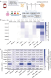
FIGURE 1
FIGURE 1
Expansion and phenotype of expanded umbilical cord blood natural killer cells (NK) with different treatments. (A) Methods of NK cell expansion with different irradiated feeder cells. (B) Heatmap showing fold change in NK cell expansion from six donors cultured with or without engineered feeder cells, normalized to wild‐type K562 feeders. (C) Heatmap showing the fold change in the percentage of NK cells expressing activating or inhibitory receptors when cultured with different feeder cells, normalized to wild‐type feeder cells. CD56+ NK cells were gated, and double‐positive percentages normalized to the wild‐type group. Panels B and C present mean ± SEM from six donors. Statistical significance was assessed using one‐way ANOVA with Tukey's multiple comparison test. NK cells co‐cultured without feeders (Feeder‐free) or with irradiated K562 derivatives: Wild‐type; 41BBL; mbIL21; 41BBL‐mbIL21; CD80; CD80‐mbIL21; CD80‐41BBL; and CD80‐41BBL‐mbIL21.
FIGURE 2
FIGURE 2
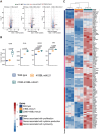
CD80‐41BBL‐mbIL21 K562 feeder cells induced the expression of genes associated with NK cell proliferation, cytokine production, and cytotoxicity. (A) Volcano plots show three pairwise comparisons: Wild‐type vs. 41BBL‐mbIL21, wild‐type vs. CD80‐41BBL‐mbIL21, and 41BBL‐mbIL21 vs. CD80‐41BBL‐mbIL21, highlighting genes linked to CD80 signaling. (B) Expression of CD80 signaling genes (CD28, FLNA, GRAP2, and VAV1) was analyzed using one‐way ANOVA with Tukey's test. (C) Heatmap showing gene expression across three groups: Proliferation, cytokine production, and cytotoxicity pathways. Genes were considered differentially expressed if the FDR‐adjusted p‐value was < 0.05 and |log2FC| ≥ 1. Panels B and C present mean ± SEM from three donors across three experiments. Statistical significance was determined by one‐way ANOVA with Tukey's multiple comparisons. NK cells were co‐cultured with irradiated K562 derivatives: Wild‐type, 41BBL‐mbIL21, and CD80‐41BBL‐mbIL21.
FIGURE 3
FIGURE 3
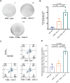
CD80‐41BBL‐mbIL21 K562 feeder cells enhanced the effector functions of NK cells. (A) Representative ELISpot images showing IFNγ production by NK cells expanded with the indicated K562 feeder cells. (B) Box plot showing quantification of IFNγ spots by NK cells expanded from the indicated K562 cells. (C) FACS plots measuring the cytolytic activity of NK cells expanded with the indicated K562 cells and co‐cultured with MK‐542 cells. (D) Box plot showing the cytotoxic index of NK cells from the three tested groups. Data in B and D are from three experiments with three donors. Statistical significance was assessed by two‐way ANOVA with Tukey's multiple comparisons. NK cells were co‐cultured with irradiated K562 derivatives: Wild‐type, 41BBL‐mbIL21, and CD80‐41BBL‐mbIL21.
FIGURE 4
FIGURE 4
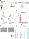
Transduction rate and cytotoxicity of CAR‐NK cells. (A) Illustration of the CAR construct expressing CEA. (B) Transduction scheme of CAR construct into NK cells using BaEV, BaEV‐VSV‐G, and VSV‐G lentiviral systems. (C) Representative FACS histograms of NK cells transduced with the indicated lentiviruses. (D) Percentage of GFP+ NK cells assessed in three different donors 5 days after transduction. (E) Microscopic observation of NK cell cytotoxic effects on MKN‐45 cells. Red arrows indicate MKN‐45 cells with damaged morphology. UTD: Untransduced NK cells; BaEV: CAR‐NK cells generated with BaEV lentivirus vector. Scale bar = 200 μm. (F) Cytotoxicity of NK cells (CCK‐8 assay) after a 4‐h co‐culture with MKN‐45 cells at different effector‐to‐target cell ratios. UTD NK: Untransduced NK cells. Mock NK: NK cells with empty vector. CAR NK: NK cells expressing CAR. Statistical significance in D and F was determined using one‐way and two‐way ANOVA, respectively, with Tukey's post hoc test.
FIGURE 5
FIGURE 5
Cytotoxicity of CAR‐NK and CAR‐T cells in 2D culture and spheroid models. (A) Microscopic observation of NK or T cell cytotoxic effects on MKN‐45 cells after 48‐h co‐culture in 2D culture. Scale bar = 200 μm. (B) Cytotoxicity index of CAR‐NK/T cells measured after 48‐h co‐culture with MKN‐45 cells. (C) Experimental procedure to assess the cytotoxicity of CAR‐NK and CAR‐T cells in cancer spheroids. (D) Confocal images show CAR‐NK/T cell infiltration (green) into 3D cancer spheroids stained with DAPI (blue) after 48 h of co‐culture. Images represent three experiments with three donors. Scale bar: 500 μm. (E) Quantification of CAR‐NK and CAR‐T cell infiltration in 3D cancer spheroids. (F) Cell death percentages in 3D cancer spheroids treated with CAR‐NK/T cells. UTD: Untransduced; CAR: Chimeric antigen receptor. Data in B, E, and F are Mean ± SEM. Significance was analyzed using two‐way ANOVA.

References

    1. Abel A. M., Yang C., Thakar M. S., and Malarkannan S., “Natural Killer Cells: Development, Maturation, and Clinical Utilization,” Frontiers in Immunology 9 (2018): 1869. - PMC - PubMed
    1. Wolf N. K., Kissiov D. U., and Raulet D. H., “Roles of Natural Killer Cells in Immunity to Cancer, and Applications to Immunotherapy,” Nature Reviews. Immunology 23, no. 2 (2023): 90–105. - PubMed
    1. Berrien‐Elliott M. M., Jacobs M. T., and Fehniger T. A., “Allogeneic Natural Killer Cell Therapy,” Blood 141, no. 8 (2023): 856–868. - PMC - PubMed
    1. Lamers‐Kok N., Panella D., Georgoudaki A. M., et al., “Natural Killer Cells in Clinical Development as Non‐Engineered, Engineered, and Combination Therapies,” Journal of Hematology & Oncology 15, no. 1 (2022): 164. - PMC - PubMed
    1. Tong L., Jiménez‐Cortegana C., Tay A. H. M., Wickström S., Galluzzi L., and Lundqvist A., “NK Cells and Solid Tumors: Therapeutic Potential and Persisting Obstacles,” Molecular Cancer 21, no. 1 (2022): 206. - PMC - PubMed

MeSH terms