. 2025 May;35(3):e70014.
doi: 10.1002/hipo.70014.

Transient DREADD Manipulation of the Dorsal Dentate Gyrus in Rats Impairs Initial Learning of Place-Outcome Associations

Affiliations

Transient DREADD Manipulation of the Dorsal Dentate Gyrus in Rats Impairs Initial Learning of Place-Outcome Associations

J Lim et al. Hippocampus. 2025 May.

Abstract

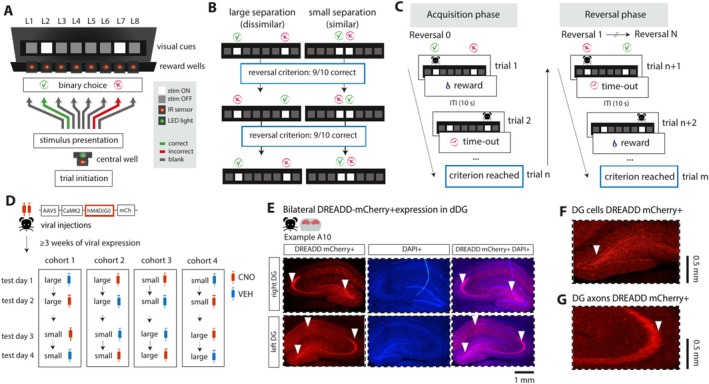
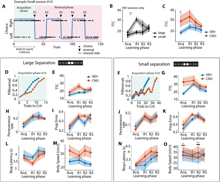
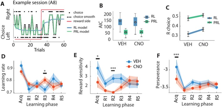
The dentate gyrus subfield of the hippocampus is thought to be critically involved in the disambiguation of similar episodic experiences and places in a context-dependent manner. However, most empirical evidence has come from lesion and gene knock-out studies in rodents, in which the dentate gyrus is permanently perturbed and compensation of affected functions via other areas within the memory circuit could take place. The acute and causal role of the dentate gyrus herein remains therefore elusive. The present study aimed to investigate the acute role of the dorsal dentate gyrus in disambiguation learning using reversible inhibitory DREADDs. Rats were trained on a location discrimination task and learned to discriminate between a rewarded and unrewarded location with either small (similar condition) or large (dissimilar condition) separation. Reward contingencies switched after applying a reversal rule, allowing us to track the temporal engagement of the dentate gyrus during the task. Bilateral DREADD modulation of the dentate gyrus impaired the initial acquisition learning of place-reward associations, but performance rapidly recovered to baseline levels within the same session. Modeling of the behavioral patterns revealed that reward sensitivity and alternation behavior were temporally associated with the DG-dependent impairment during acquisition learning. Our study thus provides novel evidence that the dorsal dentate gyrus is acutely engaged during the initial acquisition learning of place-reward associations.

Keywords: DREADDs; dentate gyrus; episodic memory; hippocampus; place‐reward associations; rats.


Figures

FIGURE 1
FIGURE 1
Task design of location discrimination task with chemogenetic silencing of dorsal dentate gyrus. (A) Schematic overview of operant box with the CS+ and CS− visual cues (white squares) presented on a black monitor screen. After trial initiation at the central well, animals made a binary choice (left or right) with a nose poke in the corresponding cued nose poke well. (B) Cued stimuli were presented with a large separation or small separation. After 9/10 consecutive correct choices, the reward contingencies switched. (C) Schematic illustration of the learning phases of the task. At acquisition, animals identified the rewarded location by trialanderror with positive (sucrose reward) and negative (time‐out) feedback. In the reversal phase, animals re‐learnt to identify the reversed target locations by adapting their choice behavior. (D) Chemogenetic targeting of the DG. Animals were bilaterally injected in the dDG with DREADD‐mCherry (N = 20). At testing, animals were injected with either CNO or saline (VEH) 30 min. before testing in a balanced within‐subject design. (E) Histological verification of an example animal (A10) with bilateral DREADD‐mCherry+ expression in dDG (left panel), DAPI expression (middle panel) and overlaid (right panel). White triangles indicate subregions of the hippocampus expressing DREADDs. (F) Close up of DREADD‐mCherry+ expression in the hilus and granule cell layer of the dDG, and (G) DG‐projecting axons in CA3.
FIGURE 2
FIGURE 2
Task behavior during location discrimination task for CNO and control treatment conditions. (A) Example choice behavior for a representative animal (A10) during a Small session with CNO treatment. Black dots indicate left‐ and right‐ward choices. The blue line indicates the rewarded side (left or right). Black solid triangles indicate when the reversal criterion was reached and reward contingencies reversed (reversal 1 as R1, reversal 2 as R2, etc.). (B) Trials to reach criterion (TTC) for baseline (VEH) treatment across Large (solid line) and Small (dotted line) sessions (N = 11). (C) TTC pooled across Small and Large sessions for animals with VEH (blue line) and CNO (orange line) treatment for each learning phase. In all subsequent panels, left‐side panels represent Large sessions (panels D, E, H, I, L, M) and right‐side panels represent Small sessions (panels F, G, J, K, N, O). (D, F) Example session showing the cumulative reversal probability across trials for the same animal (A10) as panel A with saline (VEH, blue line) and CNO treatment (orange line) during the acquisition phase. (E, G) TTC with VEH (blue line) and CNO (orange line) treatment for each learning phase. (H, J) Proportion of trials where the same choice was made as the previous trial (perseverance rate) for each learning phase. (I, K) Proportion of incorrect trials for each learning phase during Large (panel I) and Small (panel K) sessions. (L, N) Response latency (in sec) for each learning phase. (M, O) Body speed (in cm/s) during the choice epoch for each learning phase. Error bars indicate average task variables pooled across animals (mean ± SEM). Significance is indicated by *p < 0.05, **p < 0.01.
FIGURE 3
FIGURE 3
Increased choice switching associated with bilateral DG‐dependent discrimination deficit during. Acquisition learning of place reward associations. (A) Representative example session of choice behavior per trial (black dots) and smoothened across 4 trials (back dotted line) of a representative animal 8 (A8) during a Small separation session with CNO treatment. The choice trace is overlaid with the modeled behavior from the simple reinforcement learning (RL) model (blue line) and the perseverance RL (PRL) model (green line). The rewarded side is indicated with the red line (reward side switches after the animal made 9 out of 10 consecutive correct trials). (B) Median akaike information criterion (AIC) of the RL (blue square) and PRL model fits (green square) per treatment condition. (C) Per‐session correlations (R) between the observed and modeled choices per treatment condition as fitted by the RL (blue line) and PRL model (green line). (D) Median learning rate (alpha) for each learning phase with saline (VEH, blue line) or CNO (orange line) treatment. (E) Same as panel D, but for the median reward sensitivity (beta). (F) Same as panel D, but for the median perseverance rate (delta). Boxplots indicate the interquartile range (IQR) of task variables pooled across animals (median ± IQR). Error bars indicate average task variables pooled across animals (mean ± SEM). Significance is indicated by *p < 0.05, ***p < 0.001.

Similar articles

References

    1. Akaike, H. 1998. “Information Theory and an Extension of the Maximum Likelihood Principle.” In Selected Papers of Hirotugu Akaike, edited by Parzen E., Tanabe K., and Kitagawa G., 199–213. Springer.
    1. Alexander, G. E. , DeLong M. R., and Strick P. L.. 1986. “Parallel Organization of Functionally Segregated Circuits Linking Basal Ganglia and Cortex.” Annual Review of Neuroscience 9: 357–381. 10.1146/annurev.ne.09.030186.002041. - DOI - PubMed
    1. Ally, B. A. , Hussey E. P., Ko P. C., and Molitor R. J.. 2013. “Pattern Separation and Pattern Completion in Alzheimer's Disease: Evidence of Rapid Forgetting in Amnestic Mild Cognitive Impairment.” Hippocampus 23, no. 12: 1246–1258. 10.1002/hipo.22162. - DOI - PMC - PubMed
    1. Amaral, D. G. , and Witter M. P.. 1989. “The Three‐Dimensional Organization of the Hippocampal Formation: A Review of Anatomical Data.” Neuroscience 31, no. 3: 571–591. 10.1016/0306-4522(89)90424-7. - DOI - PubMed
    1. Barone, S., Jr. , Bonner M., Tandon P., McGinty J. F., and Tilson H. A.. 1992. “The Neurobiological Effects of Colchicine: Modulation by Nerve Growth Factor.” Brain Research Bulletin 28, no. 2: 265–274. 10.1016/0361-9230(92)90188-4. - DOI - PubMed

LinkOut - more resources